


Abstract
Schwannomas are peripheral nerve neurogenic tumours and although not common, laryngeal schwannomas can provide a unique challenge in diagnostic and treatment management. There are limited reports in the literature on approaches to management. A 73-year-old lady presented to the otolaryngology department after a MRI scan demonstrated an incidental right supraglottic mass. Further investigations included CT scanning and microlaryngoscopy, which only confirmed the presence of the mass with no histology diagnosis. Excision was undertaken by a laryngofissure approach and tracheostomy. Histology confirmed a benign ancient schwannoma.
INTRODUCTION
Schwannomas are one of the main subtypes of peripheral nerve neurogenic tumours [1, 2]. Schwannomas were first described by Verocay in 1908 and consists of Schwann cells [3]. These tumours can arise in patients with neurofibromatosis types 1 and 2.
Around 45% of neurogenic tumours arise in the head and neck [4, 5]. However, neurogenic laryngeal tumours are not common, comprising of ∼0.1% of all laryngeal tumours. Malignant transformation is extremely rare and such tumours are known as malignant peripheral nerve sheath tumours or neurofibrosarcomas.
Schwannomas arise in peripheral nerves that contain Schwann cells. They are slow growing and are sporadic solitary lesions affecting sensory nerves more frequently than motor nerves [4]. The four variants of schwannomas are ancient, cellular, plexiform and epitheloid [5]. The common locations of laryngeal schwannoma are in the aryepiglottic folds (80%) and in the false or true vocal cords (20%). The most frequently involved nerve is the internal branch of the superior laryngeal nerve [2].
There are few published reports in the literature about laryngeal schwannomas and approaches to management. We report a first case where histological diagnosis of laryngeal schwannoma was made through an incidental finding of a laryngeal lesion on radiological imaging and the subsequent surgical management.
CASE REPORT
A 73-year-old lady under investigation by the medical physicians was referred to the otolaryngology department after a magnetic resonance imaging (MRI) scan of the brain and cervical spine revealed an incidental finding of a mass on the right side of her larynx. A detailed history revealed a 6-week history of odynophagia and dysphagia, with no dysphonia. Examination of her nose and throat was clear with no cervical lymphadenopathy. Flexible nasoendoscopic examination showed the presence of a 2.5 cm smooth swelling on the right side of the supraglottis, arising from the area of the right aryepiglottic fold.
A computed tomography (CT) scan of the neck confirmed a heterogeneously enhancing, smooth, likely benign lesion in the supraglottic lesion extending cranially to involve the lower pharynx and the epiglottis, with significant compromise to her airway. Further assessment included a microlaryngoscopy and biopsy procedure.
The patient then underwent an excision of the right supraglottic mass via a laryngofissure approach and a tracheostomy (Figs 1 and 2). The tracheostomy was undertaken in order to secure her airway and until histological outcome was known. The operative procedure was uneventful with identification and complete excision of the lesion (Fig. 3). Postoperatively, the patient recovered well although at first, nutrition was maintained through nasogastric feeding, as swallowing was unsafe with a tracheostomy. Once histology was confirmed, decannulation of the tracheostomy occurred and the patient began to build up voice function and swallow. Histology reported a firm lobulated lesion measuring 55 × 30 × 20 mm. Microscopy revealed a well-circumscribed lesion containing spindle cells with round, regular nuclei. The spindle cells formed palisades and in some areas, had a storiform arrangement. Findings concluded a diagnosis of a benign ancient schwannoma.
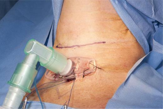
Tracheostomy prior to excision of the supraglottic mass to secure airway.
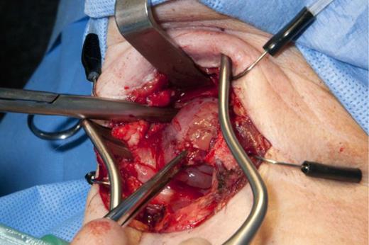
Supraglottic lesion via a laryngofissure approach.
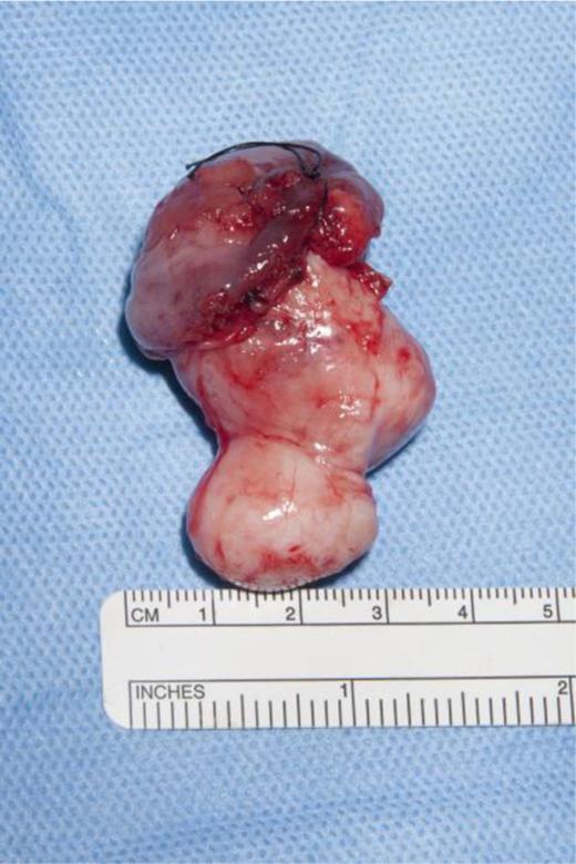
Supraglottic lesion sent for histology.
Postoperatively, 2-month follow-up showed that the patient had preserved voice quality and swallowing function with no signs of recurrence.
DISCUSSION
Schwannomas are benign, encapsulated, well-circumscribed, solitary peripheral nerve sheath tumours originating from Schwann cells. Women are twice as likely to be affected than men and it can develop at any age; however, most occur between the fifth and seventh decades [6]. It is essential to differentiate schwannomas from neurofibromas as the latter are more liable to recur and undergo malignant transformation [4].
Schwannomas classically affect nerve sheaths rather than nerve fibres and hence symptoms are determined by site and mass effect [7]. Typical symptoms of laryngeal schwannoma are hoarseness, dysphagia, dyspnoea, dysarthria, globus sensation, odynophagia, and persistent sore throat. Patients may also present with signs of airway obstruction, as in our case.
Schwannomas are classified as having three main characteristics in order to confirm diagnosis [1, 7]. (i) They arise in nerve sheaths and are encapsulated by a fibrous capsule consisting of epineurium and residual nerve fibres. (ii) They have alternating Antoni A and B areas. Antoni A areas are made up of spindle cells with twisted nuclei, indistinct cytoplasmic borders that are structured in short bundles or interlacing fascicles. Antoni B areas are less orderly and hypocellular with large, spaced vessels characteristic of schwannnomas. (iii) S-100 protein is an acidic protein found in the supporting cells of the central and peripheral nervous system and is expressed by schwannoma cells, particularly in Antoni A areas. Immunostaining for this protein acts as a main diagnostic tool. Histological findings in our case fitted these criteria to confirm a benign ancient schwannoma.
CT and MRI are useful in describing the anatomical extent of lesions and also in differentiating between malignant and benign forms. CT findings for benign laryngeal schwannoma usually include heterogenous enhancement of the lesion, no cartilage erosion and absence of infiltrative pattern [1]. Diagnosis is based on histological findings, as imaging cannot always differentiate between different tumours [8].
Surgical excision is recommended as schwannomas are radioresistant and do not recur if excised fully [1]. They are usually submucosal tumours that are extrinsic to the nerve fascicle and therefore easily separated. However, incision biopsy for schwannomas can be challenging as the capsule hinders biopsy [7].
The size and location of the lesion dictates the approach for excision. Smaller lesions can be excised endoscopically; however, larger lesions may require an external approach in the form of median and lateral thyrotomy, lateral pharyngotomy, laryngofissure or transhyoid [7]. In our case, the lesion could not be resected fully at initial microlaryngoscopy as it was difficult access; hence, the patient underwent excision of the lesion via a laryngofissure approach and a tracheostomy to protect her airway.
In summary, laryngofissure had a successful outcome in this case for a supraglottic schwannoma lesion. Surgical excision forms the mainstay of treatment for laryngeal schwannomas. The size of the lesion dictates the approach for excision and complete resection is needed to prevent recurrence. Although rare, it is important to consider a diagnosis of schwannoma with an atypical mass arising in the larynx.
CONFLICT OF INTEREST STATEMENT
None declared.
