


Abstract
Acute appendicitis is the most prevalent acute abdominal disease, but para-psoas appendix hernia is extremely rare. Here, we present the first case of a para-psoas appendix hernia, which was successfully treated with laparoscopic appendectomy and hernia repair.
Introduction
Acute appendicitis is a prevalent surgical disease with well-established diagnostic and treatment protocols, especially with the widespread adoption of laparoscopic techniques. However, some rare cases continue to pose challenges to doctors, and provide educational value to surgeons. Here, we report a case of acute appendicitis that was confirmed to be a para-psoas hernia intraoperatively, which is an extremely rare condition.
Case report
A 71-year-old female, with a body mass index (BMI) of 17.58 kg/m2, was admitted to the hospital for 2 days, due to abdominal pain. Abdominal physical assessment revealed a flat and soft abdomen, weak abdominal muscles, tender right lower quadrant, and local muscle guarding with mild rebound tenderness. Blood tests revealed white blood cells of 7.3 × 10⁹/L, with neutrophil 85.5%, and abdominal computed tomography (CT) indicated acute appendicitis and calcification beside the appendix (Fig. 1). A laparoscopy examination was conducted under general anesthesia. Intraoperatively, a hernia on the lateral side of the right psoas muscle was observed, with the appendix herniating into the hernial sac. A small amount of pus was observed in the right iliac fossa. Post-appendectomy, adhesion between the appendix and hernial sac was separated, and the appendix was then removed. The hernial sac was exposed with a 3 cm depth and a 2 × 1.5 cm neck. The hernial sac was closed with interrupted sutures (Fig. 2). Specimen anatomy revealed fecal stones in the appendix and calcification in the mesoappendix. The pathology indicated acute suppurative appendicitis with appendicular peritonitis.
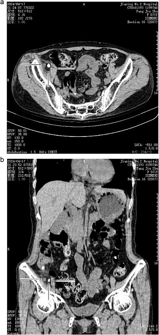
(a) Preoperative CT scan indicated acute appendicitis and calcification beside the appendix (transverse section). →: Appendix; ↑: Calcification; ←: Psoas muscle. (b) Preoperative CT scan indicated acute appendicitis and calcification beside the appendix (coronal section). →: Appendix; ↑: Calcification; ←: Psoas muscle.
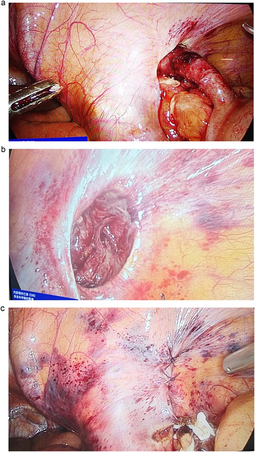
(a) Lateral hernia of the right psoas muscle, with the appendix herniating and incarcerated. (b) The hernial sac was exposed with a 3 cm depth and a 2 × 1.5 cm neck. (c) The hernial sac was closed with interrupted sutures.
Discussion
There is no reported literature on a psoas hernia with appendix as the content. The appendix, in this case, has herniated into the lateral side of the psoas muscle, causing incarceration and inflammation. Hernias that occur in space around the psoas muscle remain not uniformly defined or named, and similar case reports are rare. We tentatively name it para-psoas hernia (PPH). Modeste presented a patient with chronic groin pain who demonstrated the disease during laparoscopic surgery, with the hernia located on the lateral side of the psoas muscle and the hernia content being preperitoneal fat [1]. Badiani reported three cases of occult PPH, all of which were incidentally observed during total extraperitoneal (TEP) surgery for inguinal hernia, and the contents were all preperitoneal fat [2]. Goel presented a case of a 26-year-old patient with psoas hernia, which was discovered during TEP surgery for a left inguinal hernia, with the hernia content being preperitoneal fat [3]. Benson reported a case of a patient with a hernia of the small intestine into the left psoas muscle. Intraoperatively, the hernial sac lateral to the psoas muscle was observed, with the small intestine herniating into it. The small intestine was reduced and the hernial sac was repaired, alleviating the patient’s symptoms [4].
The hernia in this case protruded through the lateral psoas muscle, but it did not bulge on the body, making it different from a lumbar hernia. Lumbar hernia protrudes to the body through the Grynfellt triangle or the Petit triangle. However, the hernia in this patient is not a typical lumbar hernia case. The lateral psoas muscle is not a weak abdominal wall area; thus, the pathogenesis remains unclear.
In this case, further inquiry into the medical history demonstrated that the patient had undergone a hysteropexy operation >10 years ago, but the surgical process was unknown. No other history of surgery or trauma was reported; thus, the cause of the hernia remains unclear. The author holds the view that the etiology of PPH can be categorized into primary and secondary causes. Primary causes include congenital psoas muscle weakness, or age-related muscle atrophy, causing local weak areas. Secondary causes include psoas muscle injury caused by trauma, infection, or iatrogenic events. In this case, the patient reported no history of psoas surgery, trauma, or illness and exhibited a low BMI and weak muscles; thus, the author speculates that the hernia was attributed to muscle atrophy.
In this case, the depth of the hernia was approximately 3 cm and the neck was approximately 2 × 1.5 cm. The hernia could be sutured after removing the appendix. A large hernial sac requires attention in washing the wound to avoid residual infection. The patient experienced chronic abdominal pain for several years, with unclear localization, mild severity, and without relevance to eating. The pain was aggravating in the standing position but relieving in the supine position. Both gastroscopy and colonoscopy were normal. Postoperatively, the patient still had recurrent abdominal pain, with the same severity and frequency as preoperative, but the pain no longer worsened in the standing position. Therefore, the appendix hernia may be the cause of the worsening pain when standing or walking, with psoas muscle contraction squeezing the appendix, thereby causing pain.
Moreover, laparoscopic surgery should be the first choice for acute appendicitis, as comprehensive intra-abdominal exploration can determine rare conditions, such as pelvic or retroperitoneal appendices, which would cause great difficulty and risk to open surgery.
Conflict of interest statement
There is no financial and non-financial conflict of interest in the submission of this manuscript.
Funding
None declared.
