


Abstract
Trocar-site hernia is an uncommon complication of laparoscopic surgery and can be classified as early-onset, late-onset or special type. Special type hernias usually occur in the early postoperative period and result in evisceration of intra-abdominal contents through all layers of the abdominal wall without an overlying hernia sac. We present a case of special type herniation of the appendix through a 5-mm trocar site in the right iliac fossa following laparoscopic repair of a perforated duodenal ulcer. In this case, herniation occurred after removal of a drain inserted through the trocar site intraoperatively and was treated with emergent open appendicectomy. A number of patient and technical factors may be associated with an increased risk of trocar-site herniation including increasing age, elevated body mass index, increasing trocar size, longer procedure duration and absence of fascial closure. These factors must be borne in mind when planning trocar placement and number to reduce the risk of herniation.
INTRODUCTION
Trocar-site hernias are an uncommon complication of laparoscopic surgery with an incidence of 0.5% [1]. They have been classified by Tonouchi et al. as early-onset, late-onset or special type. The latter appears to be the least common type and involves complete dehiscence of all abdominal wall layers with evisceration of intra-abdominal contents through the hernial defect without an overlying hernia sac usually occurring early in the postoperative period [2]. These special type hernias usually require emergent repair in patients who may already have significant physiological derangement from previous surgery or illness and thus have the potential to cause further serious morbidity. We present a case of special type hernia following laparoscopic repair of a perforated duodenal ulcer with evisceration of the appendix through a 5-mm trocar site.
CASE REPORT
A 59-year-old female smoker with a background history of hypertension, depressive disorder and body mass index of 17 presented to the emergency department with acute onset central abdominal pain and findings consistent with a perforated peptic ulcer demonstrated on subsequent computed tomography. She underwent successful laparoscopic omental patch repair and peritoneal lavage for a perforated ulcer located anteriorly on the first part of the duodenum with resultant four quadrant peritonitis. The procedure was completed using four trocars, with a 10-mm trocar placed at the umbilicus and 5-mm trocars in the right iliac fossa, right upper quadrant and left flank. A 21-Fr Robinson drain was inserted through each of the 5-mm trocar sites with the drain in the right iliac fossa lying in the pelvis and the remaining two drains anterior and posterior to the patch repair. The patient’s postoperative course was complicated by paralytic ileus and hospital-acquired pneumonia.
The drains placed in the right iliac fossa and left flank were removed on the 10th postoperative day without immediate complication. On the 12th day postoperatively, the patient noted a swelling at the right iliac fossa trocar site. On removal of the wound dressing, the appendix and mesoappendix were noted to have herniated through the trocar site (Fig. 1). While the appendix could not be reduced, there were no signs of strangulation or apparent perforation.
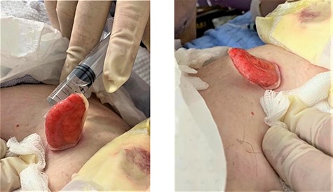
Herniation of appendix and mesoappendix through trocar site
The patient was transferred to theatre and underwent emergent open appendicectomy through extension of the trocar-site incision. The entire length of the appendix was noted to have herniated through the trocar site with the appendiceal base and caecum lying adjacent to the abdominal wall defect. There was no apparent injury to the appendix or visualized caecum. Ligation of the appendix was completed with 2-0 polyglactin with subsequent appendicectomy and inversion of the appendix stump. Continuous closure of fascial layers and skin closure with interrupted subcuticular sutures were completed.
The patient’s further postoperative course was uncomplicated and she was discharged home after a total inpatient stay of 21 days. Histopathological examination of her appendix demonstrated acute appendicitis.
DISCUSSION
Special type hernia is an exceptionally rare occurrence after laparoscopic surgery but has the potential for significant patient morbidity. While information on the incidence and characteristics of these hernias is limited to case reports, herniation of small bowel or omentum through an umbilical trocar site is most common and requires emergent repair [3]. At least two cases of evisceration of the appendix through trocar sites of 5 and 12 mm have been previously reported after elective laparoscopic procedures. In both cases, herniation also occurred through trocar sites in the right lower quadrant after removal of drains and open appendicectomy was completed in both cases [4, 5]. To our knowledge, this is the first report of a case of special-type herniation of the appendix after an emergency procedure.
There are a number of patient- and procedure-related factors, which may predispose to the development of trocar-site hernia. Patient factors associated with herniation include older age and elevated body mass index with increased intra-abdominal pressure and reduced fascial strength suggested as contributory factors. Although diabetes mellitus has been suggested as a potential risk factor, there is little evidence regarding the impact of patient comorbidity on hernia development [6, 7]. Technical factors, which may affect the incidence of trocar-site hernia, include trocar size and position, fascial closure and procedure duration. Increasing trocar size is a major risk factor for hernia development with 96% of hernias occurring at trocar sites ≥10 mm. The majority of trocar-site hernias occur in the midline or at the umbilicus, and while the single fascial layer at the linea alba may increase the risk of herniation, insertion of larger ports in these locations may also contribute [1, 8]. Fascial closure is associated with reduced incidence of trocar-site hernia, but the optimum suture type or closure method remains unclear [1]. Hernia incidence may also increase with increasing procedure duration, presumably due to increased port manipulation and stretching of fascial layers [7]. Trocar-site hernias may also occur more commonly after certain procedures with herniation more common in colorectal procedures, although the longer duration of follow-up in patients undergoing colorectal surgery may result in increased detection [8]. Interestingly, herniation at the umbilicus also appears higher in single-incision laparoscopic surgery than conventional laparoscopy, which may be a result of the larger fascial defect required for port insertion [9]. While drain insertion through trocar sites generally precludes closure of the fascial defect, its effect on the incidence of trocar-site herniation is unknown.
In the reported case, lack of fascial closure and the patient’s poor nutritional status likely contributed to hernia development. While fascial closure is not routinely completed for 5-mm trocar sites, we propose that drain insertion and manipulation may result in widening of the trocar site and poor approximation of fascial layers after drain removal. Drain insertion through trocar sites prevents fascial closure, which in itself is a risk factor for the development of trocar-site hernias. Careful consideration should therefore be given to the necessity and optimal placement of surgical drains before their insertion through trocar sites to minimize trocar-site hernias and their associated complications.
CONFLICT OF INTEREST STATEMENT
None declared.
