-
PDF
- Split View
-
Views
-
Cite
Cite
Atta Nawabi, Adam C Kahle, Clay D King, Perwaiz Nawabi, Small bowel obstruction due to retroperitoneal hernia following renal transplant: a case report, Journal of Surgical Case Reports, Volume 2020, Issue 11, November 2020, rjaa467, https://doi.org/10.1093/jscr/rjaa467
Close - Share Icon Share
Abstract
Para duodenal hernias, the most common type of retroperitoneal hernias, are thought to occur naturally from abnormal gut rotation because of fusion folds within the peritoneum. Retroperitoneal hernias are a rare postoperative complication and have not been described after renal transplantation via a retroperitoneal approach. This case report presents a 48-year-old male with intestinal obstruction after renal transplant due to herniation into the retroperitoneum via an incidentally created peritoneal defect. We suggest computed tomography with oral contrast be used in the early postoperative phase to assess for obstruction in patients with prolonged ileus of unclear etiology who have undergone retroperitoneal dissection. Small peritoneal defects should be closed during dissection. Larger, or multiple peritoneal defects should be extended to make a single, large defect to decrease the possibility of bowel herniating and becoming incarcerated.
INTRODUCTION
Early postoperative small bowel obstruction, defined as obstruction within 30 days of operation, is a relatively common postoperative complication with reported rates of occurrence between 0.7 and 24.2% [1, 2]. Retroperitoneal hernias are more rare and usually occur in natural fossa, like in Para duodenal hernias as first described by Treitz in 1857 [3]. However, there have been a handful of case reports with this complication after laparotomy due to defects created in the lateral and posterior colonic retroperitoneal attachments [4–6]. Operations performed solely in the retroperitoneal space, such as renal transplant, should theoretically avoid this complication as the intraabdominal space is not entered. However, creating small peritoneal defects is not uncommon during initial dissection while exposing the iliac vessels and developing the space for the graft. When recognized, these defects are often closed primarily with suture and experience no further complication, though there is a theoretical risk of intraabdominal contents herniating into the retroperitoneum if small defects are left unrecognized and not closed. To date, intestinal obstruction due to retroperitoneal hernia following renal transplant has not been reported in the literature.
CASE REPORT
A 48-year-old male with a history of end-stage renal disease secondary to hypertension and interstitial nephritis was admitted to our hospital to undergo a redo deceased donor renal transplantation (DDRT). His past surgical history was notable for a prior DDRT at another facility in 2011 that was complicated by rejection and, ultimately, graft failure requiring him to restart hemodialysis. The physical exam at the time of admission was unremarkable. Standard preoperative labs and imaging were obtained and were within normal limits. The patient was induced with thymoglobulin and underwent DDRT with ureteroneocystostomy and stent placement using a right renal graft that was placed in the patient’s left hemipelvis due to his prior renal transplant in his right hemipelvis.
The patient’s hospital course was notable for delayed graft function (DGF) requiring initiation of hemodialysis for hyperkalemia. Despite his DGF, he initially progressed well, however, he began to have increased abdominal pain and distention that required nasogastric tube placement and decompression on postoperative day (POD) 7. Due to a history of idiopathic gastroparesis, the department of gastroenterology was consulted, and metoclopramide was initiated. Workup by both the surgical and gastroenterology team included computed tomography (CT) imaging, esophagogastroduodenoscopy (EGD), small bowel radiograph series and endoscopic ultrasound. The patient’s CT was notable for an ileus vs. developing partial small bowel obstruction with scattered small volume pneumoperitoneum. His EGD was largely normal apart from a possible inverted duodenal diverticulum. Small bowel radiograph series demonstrated persistent obstruction, while his endoscopic ultrasound was normal. Total parenteral nutrition (TPN) was initiated on POD 13.
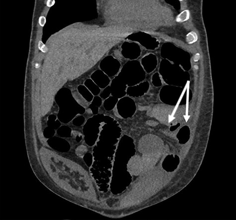
Eventually, the patient had a return of bowel function and his diet was advanced. His renal function also improved. The patient was discharged home in stable condition on POD 15 with close follow-up arranged in the outpatient transplant clinic. However, the patient was then readmitted 2 days after his discharge due to abdominal pain and inability to tolerate oral intake. He was managed conservatively, and TPN was again initiated. He underwent CT imaging, which demonstrated a partial small bowel obstruction with a transition point in the left lower quadrant of the abdomen (Fig. 1). He was operatively explored and found to have a loop of small bowel herniated into the left retroperitoneal space through a peritoneal defect. A small bowel resection with staple-assisted side-to-side enteroenterostomy was performed, and the hernia defect was closed primarily. He progressed well postoperatively and was discharged home in stable condition on POD 7.

Coronal view of non-contrast CT with arrows denoting herniated mesentery and small bowel into the retroperitoneum. Note the nonfunctional prior renal transplant in the right hemipelvis.
DISCUSSION
Hernias involving musculoaponeurotic defects, as in umbilical or inguinal hernias, are common occurrences and should be very familiar to the general surgeon. In contrast, retroperitoneal hernias are not encountered as frequently. Paraduodenal hernias, the most common type of retroperitoneal hernias, are thought to occur naturally from abnormal gut rotation because of fusion folds within the peritoneum [7].
Retroperitoneal hernias acquired later in adulthood are usually associated with traumatic injuries or at the site of prior laparotomy and occur on an even more infrequent basis. Of the cases reported in the literature, defects in the lateral and posterior colonic retroperitoneal attachments appear to be the most common source [4–6]. Hendrickson et al. [5] reported a paracolonic hernia after the development of a retroperitoneal hematoma from anticoagulation in the treatment of a pulmonary embolism, and first shed light on a previously unrecognized variant of hernia. Retroperitoneal hernias after cystectomy have also been described by Kamyab et al. [6] in the case of radical cystectomy with ileal conduit creation for bladder cancer, and by Knoepp et al. [8] after laparoscopic donor nephrectomy. Contrary to these operations, which were performed via a transperitoneal approach, we describe a retroperitoneal hernia after a renal transplant performed solely in the retroperitoneal space.
Renal transplant alone is associated with many different early postoperative complications. Most of these are related to the graft itself and include DGF, rejection or vascular and anastomotic complications such as thrombosis, stenosis, peri-transplant hematoma and anastomotic leak. However, like any open surgery, renal transplant is also prone to postoperative complications like delayed wound healing, wound infection, incisional hernia and delayed return of bowel function.
Delayed return of bowel function is most commonly due to ileus, opioid-induced constipation or gastroparesis, a complication of diabetes, which is the most common indication for renal transplant in the USA. Bowel function will usually return after conservative management with supportive care and bowel rest when necessary. However, small bowel obstruction due to a hernia should not be left off the differential following a delayed return of bowel function in a post-renal transplant patient. This can potentially occur due to herniation through a peritoneal defect created during the initial dissection and development of the retroperitoneal space.
We suggest that the workup of delayed return of bowel function after renal transplant should include CT with oral contrast to delineate whether there is true obstruction and whether the bowel has herniated into the retroperitoneal space causing incarceration. Regarding any peritoneal defects incidentally created during dissection, these can be addressed by either closure or extension of the defect. Alkhoury and Martin [4] reported an incarcerated retroperitoneal herniation after abdominoperineal resection due to a defect in the left mesocolon. They noted that there had been some controversy in the colorectal field regarding closing peritoneal defects, but due to the potential for this type of hernia, they advocated for closure. Similarly, we advocate that small defects can be closed primarily with suture. However, if slightly larger or multiple defects are created, we suggest extending the defect to make a single, large defect. This would, in essence, peritonealize the new graft and significantly decrease the chance of herniated bowel becoming incarcerated, leading to obstruction.
In conclusion, we suggest CT with oral contrast be used in the early postoperative phase to assess for obstruction in patients with prolonged ileus of unclear etiology who have undergone retroperitoneal dissection. We also underline the need for either closure or extension of peritoneal defects incidentally created during dissection to decrease the possibility of bowel herniating and becoming incarcerated.
ACKNOWLEDGEMENTS
We would like to acknowledge the Department of Surgery at the University of Kansas for their support.
CONFLICT OF INTEREST STATEMENT
The authors declare that there is no conflict of interest or financial ties regarding the publication of this case report.
FUNDING
This research did not receive any specific grant from funding agencies in the public, commercial or not-for-profit sectors.
CONSENT
Written informed consent was obtained from the patient for publication of this case report and accompanying images.
AUTHOR CONTRIBUTION
A.C.K. worked on the conceptualization, data curation, methodology, writing of original draft, review and editing. A.N. performed the validation, supervision, writing, review and editing of the manuscript. C.D.K. did the conceptualization, review and editing, and P.N. performed review and editing.


