-
PDF
- Split View
-
Views
-
Cite
Cite
Yoko Nakayama, Akio Nakagawa, Kohei Tanigawa, Taro Kawashima, Hiroyuki Monma, Iwao Kobayashi, Shiro Takase, A case of spontaneous reduction of incarcerated inguinal hernia caused by Meckel’s diverticulum, Journal of Surgical Case Reports, Volume 2025, Issue 3, March 2025, rjaf157, https://doi.org/10.1093/jscr/rjaf157
Close - Share Icon Share
Abstract
Inguinal hernias with a Meckel’s diverticulum are called Littre hernias and are rare diseases. In many cases, symptoms of incarceration or intestinal obstruction are present, and emergency surgery is generally required. However, imaging diagnosis and clinical symptoms are similar to those of incarceration of inguinal hernias, making preoperative diagnosis difficult. In most cases, diagnosis is made during emergency surgery. A 54-year-old man with abdominal pain was found to have a right-sided incarcerated inguinal hernia on abdominal computed tomography. The incarceration spontaneously reduced, although a Meckel’s diverticulum was discovered during emergency laparoscopic surgery for an inguinal hernia. Mesh repair and Meckel diverticulotomy were performed. In cases of inguinal hernias due to a Meckel’s diverticulum, it is difficult to diagnose a Meckel’s diverticulum as the cause without exploring the intestine, although laparoscopic hernia repair is useful to investigate this possibility.
Introduction
Emergency surgery due to incarcerated inguinal hernia occurs in 9% of cases [1]. If manual reduction is possible, emergency surgery can be avoided, although the success rate is approximately 60.3% [2]. Elective surgery is performed in cases of incarcerated inguinal hernias with successful manual reduction; however, exploration of the gastrointestinal tract at that time is not routine, especially in open inguinal hernia surgery. Herein, we report a case in which emergency laparoscopic surgery was performed on a patient with an incarcerated inguinal hernia that had reduced spontaneously; gastrointestinal exploration revealed that the intestine was likely to be a Meckel’s diverticulum.
Case report
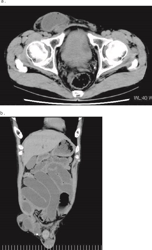
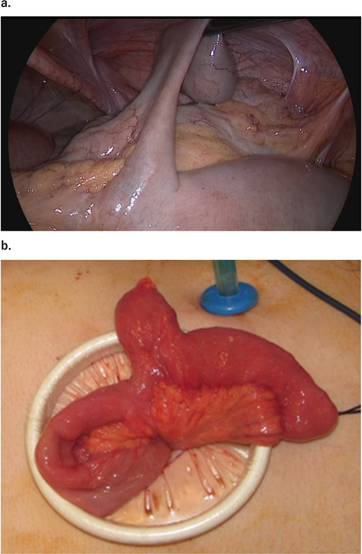
A 54-year-old, normally healthy patient had been experiencing abdominal pain for approximately 6 hours before visiting our hospital. Computed tomography (CT) revealed an incarcerated right inguinal hernia (Fig. 1); therefore, he visited our hospital. At the time of his visit, the pain had disappeared, as had the bulge in his groin. However, he still felt abdominal fullness. A repeat CT scan revealed that part of the prolapsed intestine remained in the groin. Therefore, the patient was admitted immediately and underwent emergency surgery on the same day. Laparoscopic surgery was performed under general anesthesia. Intraperitoneal findings did not confirm intestinal prolapse; therefore, right inguinal hernia repair using a transabdominal preperitoneal (TAPP) technique with placement of a 3DMax Light Mesh (Bard) was conducted. Because the patient also had an incarcerated inguinal hernia, a detailed examination of the digestive tract revealed a diverticulum on the opposite side of the mesentery, 70 cm from the terminal ileum, and dilation of the oral intestine was observed starting from this site (Fig. 2). Considering the possibility that the Meckel’s diverticulum indicated an incarcerated inguinal hernia, the intestine was elevated outside the abdominal cavity through a small abdominal incision to keep away from the mesh. The diverticulum was resected with a Powered ECHELON FLEX 3000 (ETHICON), and the stapler line was buried with a seromuscular suture to complete the surgery. Pathological examination of the diverticulum revealed ectopic gastric mucosa, although no malignancy was noted.

(a) Axial and (b) coronal contrast-enhanced CT images. The images show dilation of the intestine due to incarceration of a right inguinal hernia.

Intraoperative image. (a) Intraperitoneal observation reveals a Meckel’s diverticulum 70 cm from the Bauhin’s valve. (b) The intestinal caliber change is observed before and after the Meckel’s diverticulum.
The patient began drinking water on the day after surgery and eating on day 3 after surgery. After confirming no signs of ileus, the patient was discharged on postoperative day 8. No recurrence of the inguinal hernia has been observed 6 months post-surgery.
Discussion
An incarcerated inguinal hernia caused by Meckel’s diverticulum was reported in the 1700s by the French surgeon Alexis de Littre [3]. A Littre hernia is a rare condition, occurring in 0.09% of inguinal hernias [4] and <1% of complications of Meckel’s diverticulum [4, 5]. Littre hernias most frequently occur in the groin area, although studies have shown occurrence frequencies in the order of thigh (50%), groin (27.6%), and umbilicus (11.2%) [3]. For preoperative diagnosis, radiography is commonly used (56.1%), followed by CT (17.5%) and ultrasound (8.8%) [3], although rather than diagnosing Littre hernias preoperatively, they are generally used to support the need for emergency surgery, as Littre hernias are almost always diagnosed intraoperatively. There are a few reported cases of spontaneous reduction of an inguinal hernia caused by a Meckel’s diverticulum, as in our case. Most naturally reduced inguinal hernias are treated with elective surgery, at which point the dilation of the digestive tract is released, and surgery for the inguinal hernia is performed without exploring the digestive tract. In our case, the incarcerated hernia spontaneously decreased, and an examination was performed to check the blood flow and vitality of the released intestine, which revealed a Meckel’s diverticulum. The Meckel’s diverticulum was located on the opposite side of the mesentery of the small intestine, 70 cm from the ileocecal area. Since the caliber change of the intestine was observed before and after the Meckel’s diverticulum, it was predicted that the same part was the prolapsed intestine of the inguinal hernia.
In cases of intestinal necrosis or perforation due to a Littre hernia, intestinal resection might be avoided. In such cases, most surgeons perform tissue-only repairs to avoid mesh infection. However, clean surgery using mesh with intestinal resection, as in the present case, remains controversial. A Meckel’s diverticulum is found on the antimesenteric side, ~30–90 cm from the terminal ileum, measuring 3–6 cm in length and 2 cm in diameter [6]. A Meckel’s diverticulum is classically recognized as the cause of bleeding, ulceration, perforation, and occasionally strangulation [5]. The lifetime incidence of symptomatic Meckel’s diverticulum is 4.2%–9.0% [7], and removal completely eliminates the risk of symptoms. Park et al. reported the case of a <50-year-old male patient who had a diverticulum longer than 2 cm that contained histologically abnormal tissue and suggested that it was a symptomatic Meckel’s diverticulum [8]. In our case, the patient was over 50 years old, male, had a diverticulum over 2 cm in length, and was pathologically found to have an ectopic gastric mucosa. Therefore, it is expected that the patient presented with an incarcerated inguinal hernia and may develop symptoms in the future. The World Society of Emergency Surgery 2017 guidelines for emergency repair of complicated abdominal hernia recommend that for patients in a “clean-contaminated surgical field” needing bowel resection without fecal contamination, emergent prosthetic repair with a synthetic mesh can be performed [9]. In our case, the Meckel’s diverticulum was elevated outside the abdominal cavity to prevent contamination of the mesh, and the intestine was resected using an automatic suture machine without releasing the intestine.
Few reports of laparoscopic surgery are available for Littre hernias [3, 10], TAPP repair is useful to evaluate the intestinal tract [11] and tends to cause less postoperative pain than open hernia repair [10]. Additionally, 86.4% of Littre hernias present with symptoms of incarceration [11], and in reducible cases, elective surgery may be scheduled. If the incarcerated intestine is released using the inguinal incision method, the surgical incision may be unnecessarily long to facilitate viewing of the entire intestine. Therefore, we believe that laparoscopic hernia repair, especially TAPP, has high diagnostic and therapeutic value for released hernias of the Meckel’s diverticulum.
Author contributions
YN (drafted the manuscript), YN, HM, and IK (performed the surgeries, and all authors performed perioperative management), AN (supervised and approved the final version of this manuscript). All the authors have read and approved the final version of the manuscript.
Conflict of interest statement
The authors received no financial interest or financial support for the products or devices described in this study. The authors declare no conflicts of interest.
Funding
None declared.
Data availability
All data generated or analyzed during this study are included in this published article.
Consent for publication
Written informed consent was obtained from the patient for the publication of this case report and the accompanying images.
Ethics approval and consent to participate
Not applicable.


