


Abstract
Myelolipomas are benign tumors usually found within the adrenal gland. Approximately 50 cases of extra-adrenal myelolipomas have been reported in the literature and all are associated with additional lesions. Myelolipomas contain hematopoetic cells and adipose tissue. Most commonly, they are asymptomatic and are found incidentally on radiologic imaging. Here we report a case of an isolated intrasplenic myelolipoma as an incidental finding during the work up for myasthenia gravis in an otherwise asymptomatic man. The spleen and associated mass were excised during laparotomy and the patient had an uneventful recovery.
INTRODUCTION
Myelolipoma is a rare benign tumor composed of mature lipomatous and hemopoietic tissue, mature or immature myeloid, erythroid and megakaryocytic cells. They were first described as a finding of myeloid proliferation in the adrenal gland by Gierke in 1905 and later used in text by Oberling in 1927 [1, 2]. Myelolipomatous lesions are usually found incidentally during radiographic studies for the investigation of a patient's non-related disease [3, 4]. The discovery of myelolipoma is most commonly found with the association of the adrenal gland; however, extra-adrenal locations have been reported [5–7]. To date, splenic myelolipoma has only been described in one case in a human and in a few non-human cases [8, 9]. The authors report a case of a rare, intrasplenic myelolipoma found and treated in a human patient.
CASE REPORT
We report a case of a 64-year-old Caucasian man with a past medical history of gout, dyslipidemia, hypertension, obesity, gastroesophageal reflux disease and in previous left hemicolectomy for colon cancer nine years prior to presentation. His past surgical history includes two laparoscopic hernia repairs for incisional hernias after the hemicolectomy. He was referred to the surgery service for new diagnosis of myasthenia gravis and the consideration for possible thymectomy. The thymus was without evidence of thymoma, however, the work-up included radiographic studies demonstrating a centrally located splenic mass measuring 8 × 6.8 cm. The density of the mass was consistent with those found in soft tissue tumors (Fig. 1). A comparison CT study from 9 years previous demonstrated a smaller mass (4.7 cm) in the same anatomic location with similar density levels. There had been no prior treatments. The patient's case was presented at our multi-specialty tumor board and elective resection of the spleen was recommended due to the possibility of malignancy.
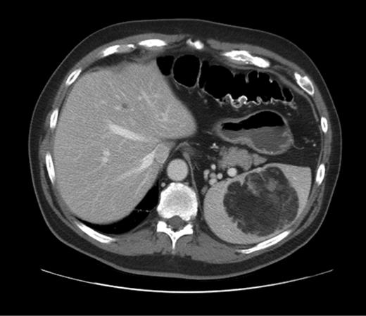
CT abdomen horizontal section with intrasplenic myelolipoma.
Exploratory laparoscopy was performed revealing significant adhesions necessitating conversion to laparotomy. An uncomplicated splenectomy was performed. Before surgery the patient was vaccinated against (Streptococcus pneumonia, Haemophilus influenza and Neisseria meningitides). Grossly, a 380-g spleen measuring 15 × 13 × 7 cm with intact splenic capsule was removed. Serial sections reveal a yellow-tan to red-tan, well-circumscribed tumor mass, measuring 10 × 10 × 7 cm. The tumor was grossly surrounded by a thin rim of dark red splenic parenchyma with splenic capsule. All sections showed a well-circumscribed area of myelolipoma completely contained within the splenic capsule (Fig. 2). The tumor consisted mainly of adipose tissue with multifocal areas of non-dysplastic hematopoietic tissue (confirmed with immunohistochemical assay positive for glycophorin and Factor VIII demonstrating erythroid elements and megakaryocyte presence). Containment within the splenic capsule was microscopically confirmed. These findings confirmed the diagnosis of intrasplenic non-neoplastic myelolipoma.
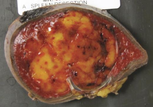
Horizontal section gross pathology of spleen with intrasplenic myelolipoma.
DISCUSSION
We describe a rare presentation of an extra-adrenal mylelolipoma in a human spleen. The etiology of myelolipoma is unclear and they are considered a type of extramedullary myeloid proliferation. O'malley et al. have proposed three hypotheses for extramedullary proliferation of hematopoietic tissue; (i) A filtration model, where immature cells are trapped by the spleen or other sites (such as in cases of acquired or functional hyposplenism) and proliferate; (ii) compromise of the marrow cavity thus hindering the appropriate numbers of marrow elements being formed or leading to increased numbers of circulating hematopoietic stem cells; (iii) abnormal signal for stem cell differentiation to hematopoietic cells and/or local effects simulating the marrow microenvironment. These signals would be through cytokine or other circulating hematopoietic growth factors [4].
The imaging, histological and pathological appearances of extra-adrenal myelolipoma are identical to those found within the adrenal gland and there appears to be no radiographic differences in these lesions from other fat containing lesions. This was demonstrated in a large case series of 76 patients, in which mylelolipoma's can be placed into four descriptive categories (i) isolated adrenal; (ii) adrenal myelolipoma with acute hemorrhage; (iii) extra-adrenal; (iv) myelolipoma associated with adrenal disease [3]. Accurate diagnosis requires tissue samples usually by fine needle aspiration and tissue biopsy methods. Radiological diagnosis can be challenging as it is difficult to differentiate this fat containing tumors from other fat containing tumors such as lipomas or sarcomas. However, histologically, these tumors are easily identified by their foci of extramedullary hematopoesis that also contain fat, are well encapsulated and contain smooth muscle as well as bone marrow cells [6].
As myelolipomas are often asymptomatic and discovered incidentally in the course of radiologic evaluation of the abdomen, they rarely present with symptoms, usually due to large size and/or abdominal pain. Patients with adrenal myelolipoma have been reported to present with symptoms such as flank pain, usually as result from tumor bulk, necrosis or spontaneous hemorrhage. The patient in this case was asymptomatic in regards to his splenic myelolipoma and his lesion was discovered incidentally. The spleen and lesion were appropriately resected in the setting of unknown pathology and demonstrates that these lesions can be managed with surgery.


