-
PDF
- Split View
-
Views
-
Cite
Cite
Saige Gitlin, Hazim Hakmi, Owen Pyke, Collin E Brathwaite, Jun Levine, Venkata Kella, Colonic herniation through the foramen of Winslow—to close the defect or not?, Journal of Surgical Case Reports, Volume 2025, Issue 1, January 2025, rjae839, https://doi.org/10.1093/jscr/rjae839
Close - Share Icon Share
Abstract
Internal herniation through the foramen of Winslow (FoW) is a rare, life-threatening diagnosis. We present a case of intestinal obstruction due to herniation of the ileum, cecum, appendix, and ascending colon through the FoW. We reduced the herniation using a small colotomy and preserved the entirety of the bowel. We closed the FoW and colotomy and secured the bowel to the lateral abdominal wall by suture cecopexy. Due to the rarity of this pathology, the literature remains unclear about the benefits of prophylactic measures, hence, we recommend continued reporting of cases and research on prophylactic measures. Further, our own treatment strategy outlined in this report may provide further insight into the misdiagnosis and management of FoW hernias.
Introduction
Internal hernias—projections of abdominal viscera through defects in the peritoneum or mesentery—constitute ⁓5.8% of small bowel obstructions [1]. Most internal hernias are acquired; however, a small subset are congenital, with hernias through the foramen of Winslow (FoW) accounting for 8% of all internal hernias [1]. Small bowel is the most common organ to herniate through the FoW, as seen in 63% of cases, followed by ascending colon (30%), and transverse colon (7%) [2].
Predisposing factors to FoW herniation are a foramen ˃3 cm, hypermobile abdominal viscera, non-retroperitoneal right colon, a large right hepatic lobe, defects of the gastro-hepatic ligament, atrophy/absence of the greater omentum, and bowel malrotations [1, 3].
Incarcerated FoW hernias require immediate surgical attention due to high risk of bowel ischemia; however, due to paucity of cases, there is no definitive best surgical management after reduction of viable of hernia contents [4].
Here, we present case of herniation of the ileum, cecum, appendix, and ascending colon through the FoW. We reduced the hernia and preserved the entirety of the bowel. We closed the defect and performed suture cecopexy to prevent recurrence.
Case
A 48-year-old female with a history of total hysterectomy, bilateral salpingo-oophorectomy, and umbilical hernia repair without mesh presented to the emergency department with chronic intermittent epigastric pain that became constant and remitting 24 h to presentation, along with one episode of non-bilious emesis. Her abdomen was diffusely tender, with severe tenderness in the epigastric region without rebound tenderness. Her vital signs were within normal limits, and laboratory workup was unremarkable.
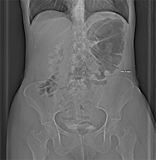
Computed tomography (CT) of the abdomen and pelvis revealed a focally dilated loop of viscera in the upper left quadrant, which was suggested to be either an internal hernia with resultant obstruction, or distention of the stomach, compatible with gastric volvulus (Fig. 1). Because of conflicting image findings, she underwent esophagogastroduodenoscopy, which was negative for gastric volvulus.

CT w/out contrast reveals focally dilated loop of transverse colon.
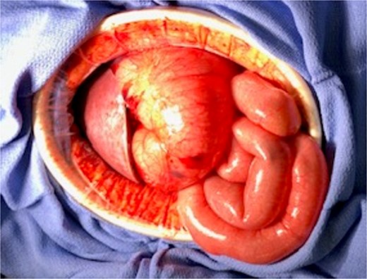
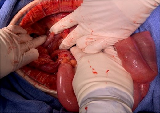
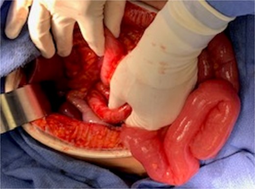
Due to the patient’s persistent pain, obstipation, and image findings, diagnostic laparoscopy was performed, which revealed a distended irreducible colon in the lesser sac. The decision was made to convert to an exploratory laparotomy, which revealed herniation of ileum, cecum, appendix, and ascending colon through the FoW causing a closed-loop obstruction (Fig. 2). Attempts at manual reduction were unsuccessful, hence a colotomy was created to decompress the air and facilitate reduction. With gentle manipulation, the colon was reduced, colotomy was closed, and then the FoW was closed with 3′0′ silk in an interrupted fashion (Fig. 3 and 4). The entire colon and bowel were healthy and viable. Afterward, a cecopexy was performed by suturing the cecum to the abdominal wall.

At laparotomy, distended loop of transverse colon with closed-loop obstruction.


The patient’s postoperative period was uncomplicated, and she was discharged on postoperative day four. She was seen in clinic in good health with complete resolution of symptoms.
Discussion
FoW hernias often present with non-specific abdominal pain and non-specific radiographic findings. Radiographic findings for FoW hernias may show bowel medial and posterior to the stomach in the lesser sac, as seen in our patient. Loops of bowel may also be visible between the liver hilum and inferior vena cava (IVC) on CT imaging [1]. In our case, the presence of viscous distention in the lesser sac was interpreted as a potential gastric volvulus. Since FoW hernias can mimic gastric volvulus on CT imaging, it is important to consider them as a differential diagnosis in patients who are preliminary identified as having gastric or cecal volvuli.
Since the first reported laparoscopic reduction of a FoW hernia in 2011, there have been a handful of laparoscopic reductions [2, 3]. To our knowledge, there are no reported robotic reductions; however, in the future robotic reduction may be considered in cases where the patient is stable to reduce the risk of a large incision. Because there has been success with the laparoscopic approach in hemodynamically stable patients, we believe a minimally invasive approach should be trialed for assessment and reduction of hernia contents when appropriate conditions are available [3]. If the hernia is too complex for laparoscopic reduction, we encourage transitioning to an open approach [3]. In our case, we were unable to visualize the extent of herniation laparoscopically and therefore switched to an open procedure.
With an open approach, we were able to reduce the hernia, preserve the entirety of the bowel, as it was non-ischemic, and secure the colon with cecopexy. In cases of ischemia, abdominal visceral resection is necessary. However, there is historical debate surrounding the necessity of abdominal visceral resection in cases of redundant abdominal viscera without ischemia. With no reported cases of recurrence with cecopexy alone, resecting non-ischemic redundant bowel may not be necessary [5]. There is little consensus regarding the necessity of cecopexy to prevent recurrences [6].
With limited data on FoW hernias, there is no consensus as to whether the hernia defect should be closed. Limited research suggests there is no significant change in recurrence at 21 months when the foramen is closed compared to left open [7]. The greatest risk when closing the FoW is the proximity to major portal structures and IVC [7]. We ultimately decided to close the foramen, guided by extensive experience with prophylactically closing acquired internal hernia spaces in gastric bypass surgeries. Specifically, a recent meta-analysis found that closure of mesenteric defects in Roux-en-Y gastric bypass may be associated with lower rates of internal herniation and reoperation [8]. However, with the overall risk of internal hernia recurrence, long term monitoring is essential in these patients. At 6 months post-surgery, there has been no reported recurrence in our patient.
Conflict of interest statement
None declared.
Funding
None declared.


