


Abstract
Uterine torsion (UT) is one of the most uncommon obstetric complications. It usually occurs in the third trimester. The confirmed diagnosis relies on > 45° rotation at the utero-cervical junction around its longitudinal axis. The etiology is unknown in 20% of cases. It might be asymptomatic in some rare cases; however, symptoms usually manifest with acute abdominal pain, fetal bradycardia, vaginal bleeding or failure of labor progress. Laparotomy is used to establish the diagnosis and the management of UT. We report a 180° uterine dextrotorsion case of an obese 24-year-old pregnant female who presented with severe abdominal pain at the 35th gestational week, which was diagnosed and managed by an emergency laparotomy. UT is a rare though serious condition and must be doubted before a suspicious clinical image. Therefore, it is suggested to avoid consecutive pregnancies and the resultant uterine rupture.
INTRODUCTION
Uterine torsion (UT) is one of the rarest obstetric complications [1–9]. The pathological torsion of the uterus is diagnosed if the rotation at the utero-cervical junction is >45° around its longitudinal axis [2–12]. It may be observed at any stage of pregnancy, but the majority of cases occur in the third trimester [3, 9]. Dextrotorsion represents two-thirds of cases, whereas levotorsion is noted in one-third [7, 8]. Many pathologies may be associated with UT: cysts, malpresentation and fetal anomalies. Approximately 20% of torsions occur without detectable causes [1, 2, 9]. The most common symptom is acute abdominal pain [1–3, 5]. However, depending on its duration and degree, UT during pregnancy is mostly symptomatic. Nevertheless, it can be asymptomatic in some rare cases. The severity and frequency of manifestations can also range from mild to severe and acute symptoms. Other clinical presentations include obstructed labor, hemorrhagic shock, gastrointestinal complaints such as nausea, vomiting and abdominal tenderness in addition to urinary complaints, including frequency, urgency, oliguria and nocturia [1, 2]. The diagnosis is confirmed mainly through a laparotomy [1, 3]. A change in position of ovarian vessels and placental site detected using Doppler and Ultrasonography, respectively, as well as the presence of an abnormal X-shaped upper vagina on Magnetic Resonance Imaging may be signs that establish an early preoperative diagnosis [9, 11]. The potential consequences of UT may include placental abruption, irregular fetal heart rate, fetal distress and fetal death, whereas chronic torsion in the early stages of pregnancy can be a cause of Intrauterine Growth Restriction [11, 12]. In all cases, prompt laparotomy with manual detorsion (MD) is considered to be an imperative treatment [1–3]. Herein, we present a case of extreme uterine dextrotorsion (UD) diagnosed in the third trimester.
CASE PRESENTATION
A 24-year-old obese female, gravida 2, para 1, presented to the Department of Obstetrics and Gynecology at the 35th gestational week (GW) with persistent abdominal pain of varying severity started at the 17th GW, not responding to analgesics. The medical history includes an umbilical hernia, insulin-controlled type I diabetes and one previous delivery through a lower segment cesarean section 3 years ago. The vaginal examination, laboratory and radiological findings were within normal limits. An emergency laparotomy was performed and revealed a 180° UD in a clockwise direction at the utero-cervical junction via Pfannenstiel incision. The left round ligament (RL) and Fallopian tube were wrapped around the lower portion of the uterus, and the left ovary was on the right side. The uterus MD in situ was unsuccessful, and therefore a transverse incision was performed on the posterior uterine wall (UW). The fetus and the placenta were delivered, with Apgar scores 8 and 9 at 1 and 5 min, respectively. The uterus was exteriorized from the abdominal cavity to correct its position by rotating it 180° around its longitudinal axis. The transverse incision on the posterior lower uterine segment has been closed (Fig. 1) without performing any incision on the uterus’s anterior surface (Fig. 2). After repositioning the uterus into the abdomen, a uterine retorsion occurred spontaneously. Due to the retorsion and to ensure the stability of the uterus in its normal position, both RL were suspended to the most proximal portion of the uterine cornu penetrating the lateral edge of the rectus abdominis muscle, and then the thread was fixed to the muscle fascia. After 6 months, the patient conceived, and the pregnancy continued without obstetrical complications until the 38th GW, despite the risk of uterine rupture (UR) because of the posterior hysterectomy. An elective cesarean section was performed, and the uterus was normal. Slight adhesions were observed and removed at the site where the RL was attached to the rectus abdominis muscle fascia. On examination, the scar on the posterior UW was normal with no adhesions. After 3 years, the fourth pregnancy occurred. The patient presented at the 33rd GW with premature labor associated with ketoacidosis which was treated immediately. The fetus was delivered, then placed in an incubator. Some adhesions were observed on the posterior uterine scar, but no recurrence of torsion was recorded.
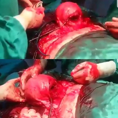
Exteriorizing the uterus intraoperatively to correct its position by rotating it 180° around its longitudinal axis. The arrow illustrates the posterior low transverse uterine incision.
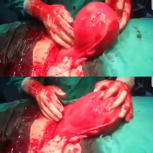
The anterior surface of the uterus obviously showing no incisions.
DISCUSSION AND CONCLUSION
When the uterus rotates around its vertical axes for >45°, it is called UT [3]. The UT angle range is between 45 and 180° [3]. However, the 180° UT has been reported in a few cases [3–5, 8, 10], (Fig. 1). Although 1/3 of the reviewed cases are levotorsion, 2/3 of most cases are Dextrotorsion [3], (Fig. 1). Although the etiology is unknown for 20% of cases [1, 2, 9], most cases are accompanied by a structural abnormality of the uterus, previous posterior lower segment incision, leiomyoma and adnexal cyst, abnormal fetal position [5]. In this case, the cause is suggested to be uterine ligament relaxation as it is re-rotated after the first attempt of correcting the UT manually and placing the uterus in the pelvic. Nevertheless, fetal malpresentation can occur [4, 6, 10] while it was normal in our case. A history of uncomplicated vaginal delivery was reported [6, 9], whereas a lower segment cesarean section history was recorded in our case. The clinical presentation could be asymptomatic [9, 10, 12] while it could be associated with fetal bradycardia, severe abdominal pain, vaginal bleeding or failure of labor progress [3]. Laparotomy approaches could exhibit different variations, as in our case where the left RL and fallopian tube stretched and wrapped around the lower portion of the uterus with the left ovary changed its position from left to the right side [2, 9]. However, ovaries, tubes and uterosacral ligaments could come together in the anterior position of the uterus [10]. Herein, laparotomy and hysterectomy on the uterus posterior face were performed; however, after the baby’s delivery, a uterus MD was done [3, 9, 12]. In conclusion, UT is a rare though serious condition and must be doubted before a suspicious clinical image. The suspension and fixation of round and broad ligaments could reduce further UT. Moreover, UR may occur with new pregnancies. Consequently, it is recommended to avoid consecutive pregnancies to prevent their severe complications.
DATA AVAILABILITY
Not applicable. All data (of the patient) generated during this study are included in this published article and its supplementary information files.
AUTHOR’S CONTRIBUTION
Marah Mansour: design of the study, data collection, data interpretation and analysis, drafting, critical revision, approval of the final manuscript.
Soulafa Alchihan: data collection, data interpretation, and analysis, critical revision, drafting, approval of the final manuscript.
Samar AlKhrait: drafting, critical revision, approval of the final manuscript.
Amro Alam Alhouda: drafting, critical revision, approval of the final manuscript.
Abdulaziz Zrik: data interpretation and analysis, critical revision, drafting, approval of the final manuscript.
Khaled Hussien: The Supervisor, patient care, drafting, critical revision, approval of the final manuscript.
ACKNOWLEDGEMENT
We are grateful to Ali Abdullatif for his help in the data collection of this manuscript.
CONFLICT OF INTEREST STATEMENT
The authors declare that they have no conflicts of interest.
FUNDING
No funding was required.
CONSENT FOR PUBLICATION
Written informed consent was obtained from the patient for publishing this case report and any accompanying images. A copy of the written consent is available for review by the Editor-in-Chief of this journal on request.
GUARANTOR
Khaled Hussien is the guarantor of this work.
