ABSTRACT
Limited evidence-based protocols exist regarding the management of venous pseudoaneurysms. Cases reports have documented surgical treatments including endovascular coil embolization or suture ligation in the setting of hemodynamic stability. Depending on the location of the venous injury, a more conservative approach to a stable pseudoaneurysm may be advantageous. This report describes a case of a patient who sustained a blunt traumatic common iliac vein pseudoaneurysm who was successfully managed non-operatively.
INTRODUCTION
Pseudoaneurysms are rare vascular lesions that result from the collection of blood between the tunica media and adventitia. Venous pseudoaneurysms occur less frequently than arterial pseudoaneurysms. The majority of pseudoaneurysms are iatrogenic, and usually secondary to manipulation of the vascular structure [1]. Other potential causes are congenital and secondary to trauma. Pseudoaneurysms are at risk for rupture causing massive exsanguination, or thrombosis, which carries the risk of pulmonary embolism [2, 3]. There are few reported cases of traumatic venous pseudoaneurysms in the literature. We present a rare case of a common iliac vein pseudoaneurysm following blunt trauma.
CASE REPORT
A 24-year-old man was brought by ambulance to our facility after having been involved in an unhelmeted motorcycle collision. He presented with sinus tachycardia up to 123 beats per minute, a Glasgow Coma Score of 15, and had an obvious deformity of the right lower extremity. His tachycardia improved with 2 L of crystalloid fluid bolus. He was stabilized in a cervical collar and a Sager traction splint on his right lower extremity and taken for CT scans of his head, cervical spine, chest, abdomen and pelvis as well as plain films of his extremities. According to our institutional protocol, he underwent CT angiogram of his neck given his skull base fracture. He was found to have a right kidney laceration with active extravasation, two large hepatic lacerations without active extravasation, a right midshaft femur fracture, bilateral distal radius fractures, a left occipital condyle fracture, a dissection of the left internal carotid artery, and pooling of IV contrast near the bifurcation of his inferior vena cava (IVC). This was concerning for a vena cava or iliac vein injury (Fig.
1). Given his hemodynamic stability, we consulted the vascular and interventional radiology teams regarding management. We decided to proceed with visceral arteriogram to control bleeding from the right kidney as well as a venogram to further delineate the suspected venous injury (Fig.
2).
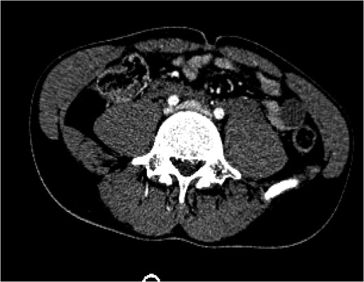
Figure 1:
A CT of the abdomen and pelvis taken on arrival (Day 1) showed a blush of contrast at the bifurcation of the common iliac veins.
The visceral arteriogram demonstrated extravasation from the right adrenal artery and superior pole of the kidney, both of which were embolized with microcoils. Post-embolization angiography demonstrated no further evidence of active extravasation. There was no injury to the aortic bifurcation. A venogram was then performed, which demonstrated a 2.8 cm pseudoaneurysm arising from the proximal portion of the left common iliac vein with involvement of the base of the IVC without active extravasation. There was no involvement of the right common iliac vein. The patient continued to be hemodynamically stable so the decision was made to observe him. After an initial hemoglobin drop from 14.3 to 6.9 g/dL within the first 48 h, he stabilized between 8 and 9 g/dL after two units of packed red blood cell transfusion and then remained hemodynamically stable throughout his stay. He was initiated on aspirin and therapeutic heparin for his carotid dissection on hospital Day 2 and underwent a cerebral angiogram and right internal carotid stent placement on hospital Day 14. A CT venogram on hospital Day 16 demonstrated a decrease in size of the pseudoaneurysm, and a stable prevertebral and pelvic retroperitoneal hematoma (Fig.
3). These were managed conservatively as the patient’s hemoglobin and hemodynamics were stable. Given the concern for intraluminal thrombus extension from the pseudoaneurysm, the decision was made to continue therapeutic anticoagulation. He was bridged to warfarin on hospital Day 17 and discharged to a rehabilitation facility on hospital Day 20 on therapeutic anticoagulation and full dose aspirin.
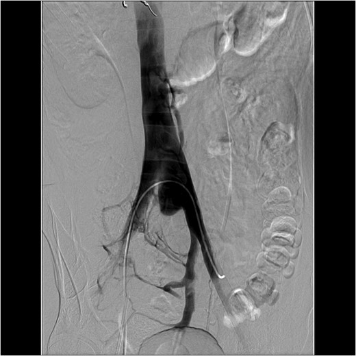
Figure 2:
An interventional radiology angiogram and venogram on Day 1 showed a 2.8 cm pseudoaneurysm in the proximal portion of the left common iliac vein. There was no involvement of the base of the IVC and no active extravasation.
DISCUSSION
Venous aneurysms, both true and false, can be either congenital or acquired. In this case, the cause of the pseudoaneurysm is hypothesized to be blunt trauma creating compression on the IVC and proximal portion of the left common iliac vein, which then led to a puncture of the inner two vessel layers. Pseudoaneurysms of internal vessels can pose a diagnostic challenge as they can rarely be detected by physical examination and are often misdiagnosed. During this case pooling of contrast was seen on CT scan, but formal diagnosis of a pseudoaneurysm was only made with a traditional venogram. As this case reinforces, venogram is the standard for diagnosing a venous pseudoaneurysm while CT angiography is adequate for surveillance of a venous pseudoaneurysm [
4].
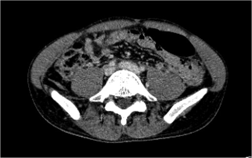
Figure 3:
A CT angiogram of the chest with and without contrast on Day 16 showed resolution of the pseudoaneurysm in the proximal left common iliac vein with clear flow at the IVC bifurcation.
Treatment for venous pseudoaneurysms should depend on the hemodynamic status of the patient as well as the location of the pseudoaneurysm. Goals should include early identification and control of exsanguination followed by recognition of and management of thrombosis. Peripheral arterial pseudoaneurysms with active blood flow, such as femoral arteries injured during catheterization, can be diagnosed with ultrasound and treated with ultrasonographically guided thrombin injection [5, 6]. Other options include endovascular coil embolization, covered stent placement or operative repair [2, 7, 8]. However, the lack of literature on venous pseudoaneurysms limits evidence-based treatment. Case reports and small series suggest endovascular coil embolization or suture ligation as potential options [4].
Our experience suggests that traumatic venous pseudoaneurysms can be observed in the hemodynamically stable patient. The proximity of the pseudoaneurysm to the IVC in our particular case made endovascular options limited. The possibility of placing a bifurcated aortic device or two covered stents was felt to be unwise due to the larger diameter of these stents leading to risk of causing additional venous injury. Surveillance imagining demonstrated a stable retroperitoneal hematoma, which we believed to be a thrombosed pseudoaneurysm. The patient has had no complications from the pseudoaneurysm as of follow-up 3 months later.
In conclusion, we report a rare case of a traumatic common iliac vein pseudoaneurysm after blunt trauma in a hemodynamically stable patient. It was diagnosed with a venogram, determined to be non-enlarging and without extravasation, observed and surveyed with CT angiography. We submit this case as a potential management option for hemodynamically stable patients with stable traumatic venous aneurysms.
ACKNOWLEDGMENTS
The authors have no conflicts of interest to report and have received no financial or material support related to this article.
REFERENCES
2Al-Damegh
SA
.
Management of
traumatic iliac vein pseudoaneurysm by transcatheter embolization .
Clin Radiol
2002
;
57
:
655
–
7
.
3Pollard
J
, Abu-Yousef
M
.
Internal jugular vein pseudoaneurysm
.
Ultrasound Q
2014
;
30
:
225
–
7
.
5Salour
M
, Dattilo
JB
, Mingloski
PM
, Brewer
WH
.
Femoral vein pseudoaneursym: uncommon complication of femoral vein puncture
.
J Ultrasound Med
1998
;
17
:
577
–
9
.
6Naimi
A
, Didier
D
, Grossholz
M
, Camenzind
E
, Chatelain
P
.
Treatment of post-coronarography femoral false aneurysm by compression, guided by Doppler echography
.
J Radiol
1996
;
77
:
247
.
7Goh
MH
, Teo
LT
, Pau
U
.
Pulmonary vein pseudoaneurysm secondary to blunt trauma: a novel management strategy
.
Ann Thorac Surg
2016
;
101
:
1197
–
200
.
8Huang
IK
, Nadarajah
M
, Teo
LT
, Ahmed
DB
, Pua
U
.
Percutaneous coil embolization of traumatic juxtacardiac right inferior pulmonary vein pseudoaneurysm
.
J Vasc Interv Radiol
2015
;
26
:
755
–
7
.
Published by Oxford University Press and JSCR Publishing Ltd. All rights reserved. © The Author 2017.
This is an Open Access article distributed under the terms of the Creative Commons Attribution Non-Commercial License (
http://creativecommons.org/licenses/by-nc/4.0/), which permits non-commercial re-use, distribution, and reproduction in any medium, provided the original work is properly cited. For commercial re-use, please contact journals.permissions@oup.com


