


Abstract
Situs inversus totalis (SIT) is a congenital condition consisting of a mirror image of transposition of the abdominal and thoracic organs occurring in about 1:5000 to 1:10 000 adults. We report on a 60-year-old male with a single colorectal liver metastasis in the Segment 7. The patients underwent a totally laparoscopic sub-segmentectomy. Intraoperative approach on a reverse posterior segment was difficult because of left-sided position of the liver. Postoperative course was uneventful and the patient was discharged after 5 days. To our knowledge, only a few cases of open liver resections in patients with SIT have been published. This is, therefore, the first case of laparoscopic liver resection for colorectal liver metastasis in a patient with SIT. We provide the readers with useful tips to perform minimally invasive liver surgery in such patients.
INTRODUCTION
Situs inversus is a congenital condition with an incidence ranging between 1:1000 and 1:10 000 characterized by left-to-right transposition of one or more normally asymmetrical organs of the body. Situs inversus totalis (SIT) consists of a mirror-image transposition of the abdominal and thoracic viscera, which occurs in about 1:5000 to 1:10 000 adults [1]. Patients affected by SIT are usually completely asymptomatic, but they have more commonly major defects which can shorten their lifespan [1].
To our knowledge, few cases of open liver resections for hepatocellular carcinoma in Japanese patients with SIT have been published [2]. In addition, laparoscopic liver resections (LLRs) in these patients have not been published by western authors. Minimally, invasive surgery can be very challenging due to the unusual anatomy.
We herein present the case of a patient with a single colorectal liver metastasis with SIT providing the readers with useful information to approach this condition.
CASE REPORT
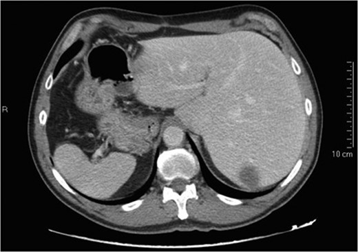
Preoperative CT scan.
Technique
The patient was placed in a right lateral position of 60°; due to the previous midline incision, we chose an open laparoscopic access with the Hasson Trocar on the pararectal line.
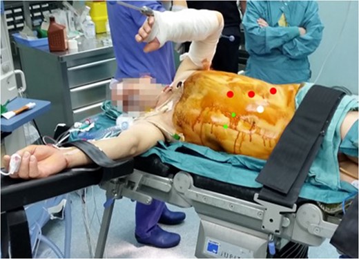
Position of the patient on the operating table and position of the trocars.
As usual, we performed an intraoperative ultrasonography with the mode of scanning was switched from normal to reverse modality; so, it was possible to have on the screen the usual appearance of the intrahepatic anatomy despite of the present specular anatomy. This trick was very useful to reach a good resection line.
The whole procedure required 240 min. We performed an intermittent (15 min with 5 min of release) Pringle maneuver for a total of 36 min.
Total blood loss was 220 ml with no blood transfusions.
The patient had the first bowel movement on postoperative day 1 and he was started on oral feeding. No peri-operative complications occurred and he was discharged home 5 days after surgery.
The specimen sent to pathology measured 7 × 3 × 5 cm with 0.9 cm clear resection margin.
At 3-month follow-up, the patient was doing well and no complications have been observed.
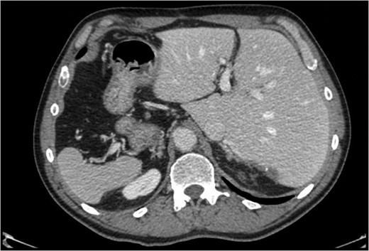
Postoperative CT scan 6 months after liver resection.
DISCUSSION AND CONCLUSION
Over the last 10 years, we observed an increase in peer-reviewed papers on LLR evaluating surgical and oncological results [3, 4].
According to Louisville statement and Morioka Consensus Conference, LLR is a feasible and safe procedure with surgical and oncological results similar to open surgery [3, 4]. Moreover, LLR seems to have a better quality of life during the first year after surgery compared with open approach [5].
At the beginning of the introduction of LLR, one of the more frequent criticisms was that, because of a more challenging approach for anatomical segmentectomies, in most cases a non-anatomical resection is performed with thinner resection margins compared with open surgery [6]. However, even in the presence of reduced surgical margins, oncological results in the short- and long-term follow-ups are the same such as open surgery especially for colorectal liver metastasis [7].
However, a non-anatomical approach seems to have the same oncological results compared with anatomic resections but with better postoperative course due to the parenchymal sparing [8].
According to the Morioka Consensus Conference ‘anatomical resections, including sectionectomy, segmentectomy and sub-segmentectomy, meant as parenchymal preserving resections of portal territories are considered complex procedures requiring identification of anatomical boundaries’ [4].
These observations are even more true for a patient with previous open surgery and an SIT with the double challenge related to the anatomic position of the liver and the posterior (7) segment involved. Another challenging maneuver was the separation of the liver from the diaphragm; the absence of any ligament made this part of the procedure very demanding to identify the correct plane from the diaphragm and the liver and minimize the bleeding.
Our routine approach for postero-lateral segments is with the patient in a supine position [9]. This supine approach allows to have a better control of the Pringle maneuver that should always be quickly available in difficult resections to perform the quickest laparotomy in the need to convert to open surgery; however, in this particular case, we chose a lateral approach with a decubitus of 60° to have a more direct access to the left-sided liver. The mirror modality of ultrasonography, the quick control of the hilum if needed and the position have simplified the surgical approach in this patient.
In conclusion, ‘difficult’ LLR resection can be safety made in experienced hands even in the presence of anatomical variations. At the same time, anatomical variations or the hypothesis of a ‘difficult’ resection should not be an absolute contraindication to LLR.
