


Abstract
Enterobius vermicularis infection is uncommon in adults, compared to children, and rarely causes significant illness. Adult infection is usually colonic in nature and found incidentally at colonoscopy. Worm migration to other tissues is rare. We here-in describe the case of a 73-year-old woman found to have biliary tree E. vermicularis—an as yet undescribed site of migration.
INTRODUCTION
Enterobius vermicularis is a common parasitic infection which usually affects children and rarely causes significant illness. Infection is most commonly confined to the intestinal tract with involvement of other systems, such as lung, liver, breast and spleen [1] less frequently seen. Of the few reports of extraintestinal E. vermicularis infestation, patients can have significant morbidity associated with infection and in some cases mortality [2]. We report the case of a 73-year-old woman who was found to have incidental biliary infestation of E. vermicularis during a laparoscopic cholecystectomy.
CASE REPORT
A 73-year-old woman presented to the Emergency Department with sudden onset, post-prandial, severe epigastric pain associated with nausea and vomiting which had started 3 h prior to presentation. She denied chest pain or shortness of breath and was well until her symptoms started. There was no radiation of the pain, nor any other associated features. She had no significant past medical history and took only anti-hypertensives for blood pressure control.
Initial examination revealed a tender epigastrium and right upper quadrant but nil else significant. Observations showed that she was afebrile, in sinus rhythm, and normotensive. Blood tests revealed a white cell count of 9.7 ×10⁹ l–1, C-reactive protein < 1 mg/l, serum amylase 72 U/l, bilirubin 8 µmol/l, alkaline phosphatase 119 U/l, ALT (SGPT) 49 U/l, AST (SGOT) 38 U/l and Gamma-GT 29 U/l.
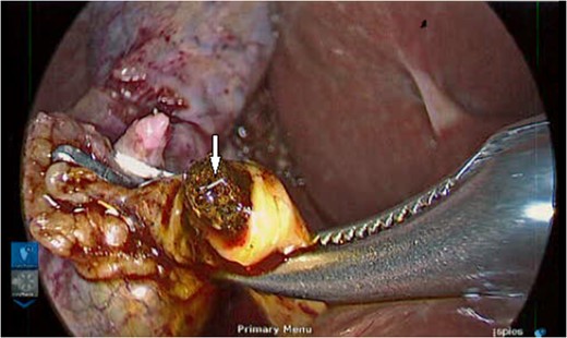
A live helminth is seen on the stone during extraction.
Although cholangiography confirmed a sub-centimetre common bile duct stone that could not be retrieved laparoscopically, the remainder of the procedure was uneventful and she recovered well from surgery to receive endoscopic retrograde cholangiopancreatography (ERCP) and stone clearance during the same admission.
When informing the patient of the intra-operative findings following the procedure, she reported that her extended family had been infected with pinworms (E. vermicularis) in the days and weeks leading up to her admission—information that had not been volunteered during admission clerk-in. She received a course of Mebendazole and following ERCP was discharged home.
DISCUSSION
Unlike the more common Billiary Ascariasis, where it is estimated up to 33% of the world's population are infected with the causative organism, Ascaris lumbricodies [3], infection of the biliary tree with E. vermicularis, more commonly known as pinworm, is rare. In contrast with the former, it is not visible on imaging and so was not anticipated intra-operatively in this case. Infection with E. vermicularis is most commonly seen in children with the majority of patients presenting with perianal pruritis. Indeed, the patient we present described her extended family, particularly grandchildren, being infected in the weeks prior to her admission, information that was not known in the peri-operative period. Given the intra-operative findings and history from the patient, this raises the likelihood of migration of the infection to the biliary tree from the intestinal tract.
Whilst usually no serious complications arise from infection with E. vermicularis, there have been cases of infection resulting in appendicitis [4] and some evidence to suggest that in adults there is a greater risk of developing this if infected [4]. To further add diagnostic uncertainty in these patients, it has also been reported that infection can mimic an acute appendicitis with no underlying inflammation being found during diagnostic laparoscopy [5]. With E. vermicularis usually being confined to the terminal ileum and large bowel, extraintestinal migration is rare. Although uncommon, ectopic enterobiasis often carries greater morbidity and in one reported case, mortality [2] due to visceral perforation and secondary abdominal sepsis.
With regard to the upper gastrointestinal tract, hepatic involvement is very rare with only five identified cases reported in the world-wide medical literature [6–10], often with patients presenting with non-specific symptoms. One reported case was initially treated as a malignant hepatic tumour and only after partial liver resection did histology confirm the underlying diagnosis [6]. Furthermore, biliary involvement is uncommon and as such we found no reports of infestation within the cystic duct or gallbladder on review of the current literature. Our case raises the possibility of biliary infestation with E. vermicularis since helminths consistent with this were clearly extracted from the cystic duct. However, unlike the cases of appendicitis secondary to infestation, given this patient's underlying gallstone disease, it is unlikely that this was involved in the aetiology of her presentation with biliary colic.
CONFLICT OF INTEREST STATEMENT
None declared.
