Abstract
A 69-year-old female with a history of bilateral total hip replacements presented with rigors, fever and sudden onset left groin pain. A pelvic X-ray showed well-fixed implants. Blood results revealed a leucocytosis (white cell count 22.3 × 10⁹ l–1) and elevated C-reactive protein (211 mg/l). Ultrasound-guided aspiration of her left hip grew Streptococcus gordonii. No source infection could be identified apart from a new chronic sinus infection in a left upper incisor. Following a discussion with the patient a 6-week course of intravenous ceftriaxone was started and was successful in normalizing her inflammatory markers. She was placed on long-term suppressive amoxicillin following this. Her suppressive antibiotic therapy was complicated by the development of a clostridium difficile infection and her antibiotics were changed to doxycycline. At 1-year follow-up, she was asymptomatic with no further episodes of groin pain or fever.
INTRODUCTION
Prosthetic joint infection (PJI) following total hip replacement (THR) occurs in 1–2% of procedures but is one of the primary reasons for prosthesis revision (1). We report the only case of Streptococcus gordonii, a member of the viridans group streptococci (VGS), causing PJI in the setting of THR.
CASE REPORT
A 69-year-old female, with a number of comorbidities, presented with an 1-day history of rigors, fever (39°C) and severe left groin pain on a background of previous bilateral cementless THRs, left THR 4 years previously and right THR 2 years previously. Her radiographs demonstrated a well-fixed implant and no periprosthetic fracture (Fig.
1). Blood results showed a leucocytosis (white cell count (WCC) 22.3 × 10
9 l
–1) and elevated C-reactive protein (CRP, 211 mg/l). Blood cultures were negative. Examination on this admission revealed a chronic sinus infection in a left upper incisor not noted on her last dental review 10 months previously. The patient described no history of gum or tooth sensitivity and pain. An orthopantomogram was done which showed multiple dental fillings but no sign of dental abscess (Fig.
2). An echocardiogram done to out rule endocarditis was normal. A computed tomography scan revealed extensive soft tissue swelling over the left hip involving the psoas muscle (Fig.
3). Ultrasound-guided aspiration was performed and 30 ml of purulent fluid was aspirated. Following this empiric antibiotics (vancomycin and gentamicin) were started. The hip aspirate grew
S. gordonii (MALDI-TOF, Bruker) that was pansensitive. Antibiotics were rationalized following consultation with the Microbiology Department. Despite a discussion in which the need for replacement of the prosthesis was had, the patient choose to initially trial conservative management with intravenous antibiotics due to her pre-existing comorbidities and wish to avoid further surgery. The risks of this course of management were explained including the ultimate need for prosthesis replacement. A 6-week course of intravenous ceftriaxone was started and was successful in normalizing her inflammatory markers (WCC 7 × 10
9 l
–1, CRP < 5 mg/l). She was placed on long-term suppressive amoxicillin following this and underwent extraction of her left upper incisor 2 weeks after the completion of her intravenous antibiotics. Unfortunately, 6 months into her treatment she developed fever, nausea and diarrhoea, and a stool sample was positive for clostridium difficile. Her antibiotics were rationalized to Doxycycline. Since then serial monthly blood tests have remained normal. At 1-year follow-up, she was asymptomatic. She had experienced no further episodes of groin pain or fever and had returned to her baseline level of mobility. Her Oxford hip score was 43 out of 48. Blood results showed a WCC of 6.7 × 10
9 l
–1 and a CRP of 11 mg/l.
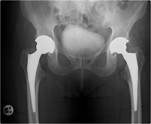
Figure 1:
X-ray pelvis—showing bilateral well-fixed THRs.
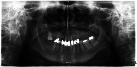
Figure 2:
Orthopantomogram—showing dental implants and fillings.
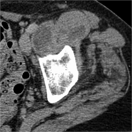
Figure 3:
Computed tomography scan showing left iliopsoas involvement and heterogeneity, with a well-defined intramuscular hypoattenuation typical for abscess formation.
DISCUSSION
PJI occurs via direct inoculation, contiguous or haematogenous spread. Late onset infections (>12 months after surgery) are predominantly due to haematogenous seeding of virulent pathogens at another site (urinary tract, soft tissue infection). PJI is primarily caused by staphylococci with only a small percentage caused by streptococci, <10% [1, 2]. Streptococcus gordonii, a member of the Streptococcus Sanguinis group, a subdivision of VGS, is one of the microbiota of the oral cavity that contributes to plaque formation. It is recognized in the setting of bacterial endocarditis [3], but has only recently been described in the literature as causing PJI, in the setting of total knee replacement reported in 2015 [4].
The reasons for the emergence of S. gordonii in PJI are not certain. One reason may be due to improved diagnostic technologies. Historically, the automated identification systems for VGS have been problematic due to changing taxonomy and addition of new species. Another reason for this is that all species are not represented in identification databases [5, 6]. In our institution, we have moved from API Strep (Biomérieux) identification system to MALDI-TOF (Bruker) identification allowing us now to accurately identify members of the streptococcus genus. Exact identification of the streptococcal organism is important, if further aspirations are required in order to reduce confusion that may result if different organisms are detected.
Treatment of PJI is a complex and challenging practice. Guidelines advise debridement, antibiotics and implant retention for patients who present with a well-fixed implant and no sinus tract within 30 days of implantation [7, 8]. For other patient's, replacement of the prosthesis or resection arthroplasty should be performed [9]. Rarely, medical therapy alone may be considered where medical comorbidities preclude against the above or where patient's autonomy must be respected. In our case, the patient was reluctant to undergo any further surgery. Little research exists regarding the treatment of PJI with antibiotics alone and the role of suppressive long-term antibiotics. Treatment failure with suppressive antibiotics is most commonly seen with staphylococci bacteria [10]. Chronic suppressive antibiotics are not without their risks, as seen in our case. It is important that fully informed consent is achieved prior to beginning the above treatment.
While the rate of PJI is low, the increasing demand for joint replacement will cause a considerable burden of infection in the future. Recognizing and managing PJI promptly is vital in reducing patient morbidity and mortality as well as costs [11, 12]. PJI will impose increasing challenges due to an ageing population, comorbidities and the emergence of increased antimicrobial resistance. Our case highlights a previously undescribed pathogen in late PJI post-THR.
CONFLICT OF INTEREST STATEMENT
None declared.
REFERENCES
1Tande
AJ
, Patel
R
.
Prosthetic Joint Infection
.
Clin Microbiol Rev
2014
;
27
:
302
–
45
.
3Murdoch
DR
, Corey
GR
, Hoen
B
, Miró
JM
, Fowler
VG
Jr, Bayer
AS
, et al. .
Clinical presentation, etiology, and outcome of infective endocarditis in the 21st century: the International Collaboration on Endocarditis-Prospective Cohort Study
.
Arch Intern Med
2009
;
169
:
463
–
73
.
4Klein
R
, Dababneh
AS
, Palraj
BR
.
Streptococcus gordonii prosthetic joint infection in the setting of vigorous dental flossing
.
BMJ Case Rep
2015
Aug 11;
2015
.
5Gavin
PJ
, Warren
JR
, Obias
AA
, Collins
SM
, Peterson
LR
.
Evaluation of the Vitek 2 system for rapid identification of clinical isolates of gram-negative bacilli and members of the family Streptococcaceae
.
Eur J Clin Microbiol Infect Dis
2002
;
21
:
869
–
74
.
6Murray
PR
.
Matric assisted laser desorption ionization time-of-flight mass spectrometry: usefulness for taxonomy and epidemiology
.
Clin Microbiol Infect
2010
;
16
:
1626
–
30
.
7Osmon
DR
, Berbari
EF
, Berendt
AR
, Lew
D
, Zimmerli
W
, Steckelberg
JM
, et al. .
Diagnosis and management of prosthetic joint infection: clinical practice guidelines by the Infectious Diseases Society of America
.
Clin Infect Dis
2013
;
56
:
e1
–
25
.
8Minassian
AM
, Osmon
DR
, Berendt
AR
.
Clinical guidelines in the management of prosthetic joint infection
.
J Antimicrob Chemother
2014
;
69
:
29
–
35
.
9Leone
S
, Borre
S
, Monforte
AD
, Mordente
G
, Petrosillo
N
, Signore
A
, et al. .
Consensus document on controversial issues in the diagnosis and treatment of prosthetic joint infections
.
Int J infect Dis
2010
;
14
:
67
–
77
.
10Prendki
V
, Zeller
V
, Passeron
D
, Desplaces
N
, Mamoudy
P
, Stirnemann
J
, et al. .
Outcome of patients over 80 years of age on prolonged suppressive antibiotic therapy for at least 6 months for prosthetic join infection
.
Int J Infect Dis
2014
;
29
:
184
–
9
.
11Cahill
JL
, Shadbolt
B
, Scarvell
JM
, Smith
PN
.
Quality of life after infection in total joint replacement
.
J Orthop Surg (Hong Kong)
2008
;
16
:
58
–
65
.
12Coello
R
, Charlett
A
, Wilson
J
, Ward
V
, Pearson
A
, Borriello
P
.
Adverse impact of surgical site infections in English hospitals
.
J Hosp Infect
2005
;
60
:
93
–
103
.
Published by Oxford University Press and JSCR Publishing Ltd. All rights reserved. © The Author 2017.
This is an Open Access article distributed under the terms of the Creative Commons Attribution Non-Commercial License (
http://creativecommons.org/licenses/by-nc/4.0/), which permits non-commercial re-use, distribution, and reproduction in any medium, provided the original work is properly cited. For commercial re-use, please contact journals.permissions@oup.com


