


Abstract
Peristomal pyoderma gangrenosum (PPG) is a rare subtype of pyoderma gangrenosum that is difficult to diagnose and treat. It is characterized by the rapid progression of painful necrotic ulcer surrounding an area of abdominal stoma. It is almost exclusively associated with inflammatory bowel disease even after bowel surgery and is associated with significant morbidity. Diagnosis of pyoderma gangrenosum is based on exclusion of other disorders replicating some of its clinical features and histopathological evidence.
This is a case report of a 56-year-old lady with rheumatoid arthritis who presented with rapidly progressing abdominal ulcer 8 months after a Hartmanns procedure for perforated diverticulitis. The ulcer had formed a large cavity causing faecal filling in the dependent defect. The other causes of ulcer were excluded with negative histopathology, negative polymerase chain reaction for Mycobacterium ulcerans and negative acid fast bacillus (AFB) test. She was diagnosed with PPG which is routinely treated medically due to risk of setting off a second focus of pyoderma if surgically intervened. However due to increased risk of faecal peritonitis, it was decided to proceed with surgical debridement. This article will discuss the case in more detail and briefly discuss diagnosis and treatment options for PPG.
INTRODUCTION
Pyoderma gangrenosum is among a group of idiopathic neutrophilic dermatosis and is considered a reaction to an internal disease or condition such as inflammatory bowel disease (both ulcerative and Crohn's disease), rheumatoid arthritis and paraproteinaemia [1]. The characteristic lesion is ulceration with a well-defined undermined violaceous border [2].
Peristomal pyoderma gangrenosum (PPG) is a rare subtype of pyoderma gangrenosum that is difficult to diagnose and treat, characterized by the rapid progression of painful necrotic ulcer surrounding an area of abdominal stoma [3]. Diagnosis of pyoderma gangrenosum is based on exclusion of other disorders replicating some of its clinical features and histopathological evidence [4]. Histopathology is usually nonspecific and characterized by dermal infiltration of neutrophils but helps to exclude other causes of ulceration that may be bacterial or mycobacterial infections, malignancy, vasculitis, and vascular insufficiency [5]. This case report discusses the treatment of a patient with rheumatoid arthritis who developed pyoderma gangrenosum on a background of recent abdominal surgery.
CASE REPORT
Eight months following Hartmanns procedure for perforated diverticulitis, a 56-year-old lady with a history of severe rheumatoid arthritis presented with a 3 cm large painful, rapidly progressing ulcer surrounding her colostomy site. The patient had consulted the dermatologist 1 month prior to presentation to the surgical unit and was commenced on twice weekly etanercept injections, intralesional triamcinolone, prednisolone, and tacrolimus ointment. She was also on methotrexate and leflunamide. Testing of the ulcer included polymerase chain reaction for M. ulcerans, herpes multiplex and AFB culture—all of which were negative; site MCS showed evidence of mixed enteric bacteria. After excluding other causes of ulcer and based on clinical presentation she was diagnosed with a PPG (Fig. 1). The dermatologist concerned about the risk of koebnerizing a second focus of pyoderma, recommended conservative management till pyoderma was completely turned off.
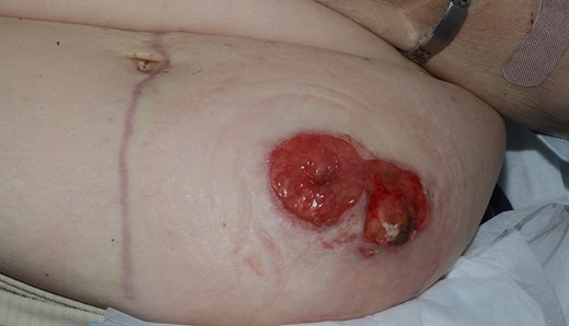
Peristomal pyoderma gangrenosum with presence of necrotizing ulcer near stoma site prior to surgery.
The rapidly progressing peristomal ulcer, however, had progressed to form a large cavity with prolapsing granulation tissue and faecal filling in the dependent defect. Due to the increased risk of bowel obstruction and faecal peritonitis, a decision was made to surgically debride the ulcer. She was given IVIG and IV methylprednisolone 3 days prior to surgery and advised regular immunosuppressants and tacrolimus ointment over wound site post operatively.
She had revision of end colostomy with debridement of peristomal ulcer and repair of parastomal hernia. Operative findings showed evidence of omentum attached to transverse colon prolapsing through ulcer. Histological results showed evidence of thick neutrophilic and fibrinous surface exudates with an underlying band of fibrous scar; and presence of reactive fat necrosis. Post operatively the patient recovered well with no further surgical complications. At the time of writing the case report it had been 12 months post surgery and patient had no evidence of peristomal ulcer recurrence (Fig. 2).
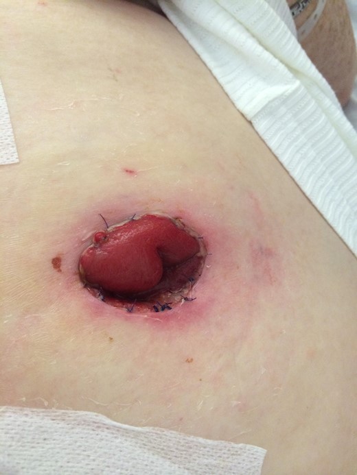
Healed site of peristomal pyoderma gangrenosum with no evidence of recurrence 12 months post op.
DISCUSSION
PPG is a disabling condition and is often challenging to treat. Approximately 25% of cases are caused by incidental or iatrogenic trauma with 14% of cases of post surgical PPG precipitated by abdominal surgery [6]. In this case PPG was associated with rheumatoid arthritis and occurred within 8 months of abdominal surgery. According to some studies, the time of onset of PPG after surgery can range from 2 months to 25 years [7].
There seems to be a scarcity of randomized controlled trials assessing the optimum treatment for this condition. Although TNF blockers, prednisolone, cyclosporine, IVIG, or plasmaphoresis and methotrexate have emerged as favourable treatment options [8], PPG can often be intractable to these conventional therapies. Surgical interventions including ulcer debridement and grafting are usually avoided during active phase of disease due to the high risk of triggering the formation of a new lesion at the surgical site [9]. However, there have been a few reports, similar to our case, of successful PPG wound management with surgical intervention. Surgical treatment is best performed in selective patients in whom medical management would risk patient mortality. These patients require systemic immunosuppressive therapy prior to surgery to avoid risk of recurrence [10].
Conflict of Interest Statement
None declared.
