


Abstract
A 31-year-old patient with obstructive voiding symptoms and apareunia in the setting of Type III female genital mutilation/cutting (FGM/C) is presented. The patient underwent ambulatory clinic defibulation to relieve her symptoms. FGM has been shown to have serious immediate complications and many chronic complications that greatly impact patients’ lives. Several case series have been published describing center-specific experience with defibulation procedures for Type III FGM/C. Here, we present the treatment of a patient with Type III FGM/C in an ambulatory urology clinic in the United States.
Introduction
A 31-year-old otherwise healthy woman presented with the primary complaint of obstructive voiding symptoms and dysmenorrhea. She reported bothersome obstructive voiding symptoms with urinary straining, a weak spray with dribbling rather than a stream of urine. She also experienced dysmenorrhea with difficulty passing menses and apareunia. She had recently married and was interested in proceeding with sexual intercourse and conception, but was very concerned about the ability to achieve penetration due to a nearly obliterated vagina.
The patient had undergone female genital mutilation/cutting (FGM/C) at the age of 10 in her home country of Morocco. The procedure was performed within the local mosque by a religious affiliate without medical training and without any form of anesthesia. She recalled excessive bleeding and intermittent urinary retention for 3 days after the procedure. The physical and psychological sequelae of the procedure were profound and were also leading to new stress within her recent marriage.
Since her move to the United States, she had presented for assessment and treatment to medical practitioners (including urologists) in two centers who were uncomfortable providing treatment. In addition, her lack of insurance and inability to pay out of pocket limited her access to health care.
Case Report
Physical examination in the dorsal lithotomy position revealed complete fusion of the labia majora with a small osteum below the fused labial bodies leading to a very small posterior introitus (<1 cm). The clitoris was not visible and speculum examination was impossible. The urethra was not visible on initial evaluation, but the patient acknowledged urinary spraying at a level above and below the fused labial bodies (Fig. 1). Probing of the fusion site with a narrow swab demonstrated communication between the upper and lower fossa behind the labial fusion (Fig. 2). The remainder of the pelvic examination was normal.
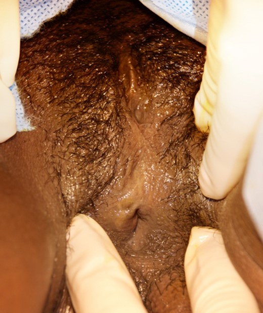
Initial clinical evaluation at presentation demonstrating fusion of the labial bodies.
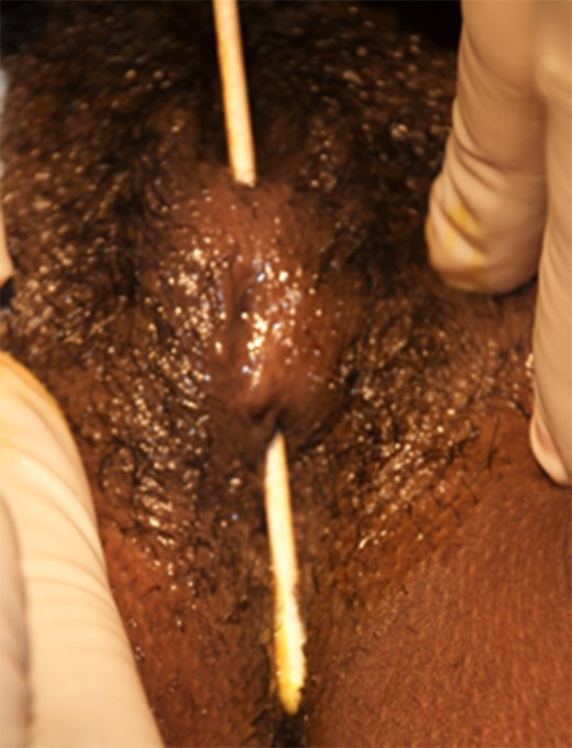
Clinical evaluation at presentation demonstrating fusion of the labial bodies. A simple swab demonstrates communication behind the labial fusion.
The patient requested defibulation (reconstructive surgery of the infibulated scar) in-office following discussion of risks and possible complications. Management in the operating room was offered and encouraged, but she declined this option due to lack of insurance. The fused labia were infiltrated with 0.5% Marcaine with epinephrine and a cotton tip applicator was used to palpate the introitus to locate a tract behind the labia (Fig. 2). Hemostats were placed across the bridge of tissue to cause crush and assist with hemostasis. Metzenbaum scissors were used to divide the labia across the scar in the plane created by the hemostats over the swab to protect the structures behind the probe. The two free edges of the labia were then reapproximated with absorbable suture. Examination after defibulation showed an intact clitoris, a patent urethral meatus and normal vaginal tissue (Fig. 3). Bimanual exam was also normal.
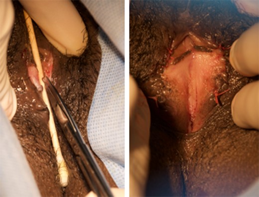
Defibulation procedure: incision along the tract of labial fusion (left) and open vaginal introitus following defibulation (right).
Follow-up at 6 weeks revealed a well-healed incision with no evidence of adhesion. The patient had commenced sexual activity without difficulty and her urinary complaints had also completely resolved. She and her husband expressed satisfaction with the procedure and the results.
Discussion
FGM/C, or female circumcision, is defined by the World Health Organization as ‘partial or total removal of the external female genitalia or other injury to the female genital organs’ [1]. Worldwide, there are an estimated 125 million girls and women living with FGM/C with an estimated 228 000 cases of FGM/C in the United States [1, 2]. The WHO classifies FGM/C into four categories: excision of the prepuce, with or without partial or complete clitoral excision (Type I); clitoral excision with partial or total labia minora excision (Type II); partial to complete excision of external genitalia and stitching/narrowing of the vaginal opening known as infibulation (Type III); otherwise unclassified including different cultural practices (Type IV).
FGM/C has been associated with negative health outcomes in multiple studies and reviews [3–5]. Immediate complications include bleeding, urinary retention, wound infection, sepsis and death. Long-term complications include dysmenorrhea, dyspareunia, recurrent vaginal and urinary tract infections, infertility, difficult labor and delivery and sexual dysfunction [3–5]. Only one study has focused on lower urinary tract symptoms (LUTS) following FGM/C and concluded that all variants of LUTS were significantly higher in those with Type II and Type III FGM/C than in controls or those with Type I FGM/C [6]. In fact, women who undergo Type III FGM/C are at the highest risk for immediate and long-term complications [7].
In many areas where FGM/C is common, women have a defibulation procedure performed immediately after marriage to facilitate consummation and childbirth. However, for women now living in the West, there is difficulty in obtaining such services because they are often performed by the same community and religious affiliates who performed the circumcision in their home country [8]. We publish our experience treating Type III FGM/C in a controlled environment in the clinic setting to show that it is a viable and potentially preferable treatment in this population.
Three series detailing the treatment, or defibulation, of women with Type III FGM/C have been published. McCaffrey et al. detailed their experience with the treatment of 50 women [8]. They concluded that defibulation by cold knife cut under general anesthesia, or spinal anesthesia for pregnant patients, is ideal and is ideally performed in the antenatal period. Penna et al. detailed defibulation of 25 patients utilizing a carbon dioxide laser with no complications and concluded that it is a suitable alternative treatment [9]. All of the patients in this study were treated in the outpatient setting with local infiltration of Mepivacaine. Finally, Nour et al. published a retrospective analysis of 40 patients who underwent defibulation at Brigham and Women's Hospital [10]. These were performed under general and local anesthesia and follow-up interviews revealed high satisfaction rates among both patients.
Defibulation has shown to be a safe and effective way to treat women with Type III FGM/C and can be offered in a controlled ambulatory setting with appropriate planning and consent. We present this case report of the management of a severe case of Type III FGM/C to demonstrate treatment options requiring minimal infrastructure.
Abbreviations
(FGM/C): female genital mutilation/cutting
(LUTS): lower urinary tract symptoms
Conflict of interest statement
None declared.
Funding
Dr Khavari is supported by NIH grant K12 DK0083014, the Multidisciplinary K12 Urologic Research (KURe) Career Program.
