


Abstract
Spontaneous dissection of the coronary arteries is a rare disease with a wide range of clinical presentations ranging from angina to myocardial infarction (MI); its pathophysiology has not yet been fully established. In this paper, we present the case of a 31-year-old male with an acute coronary syndrome. The initial results of the electrocardiogram and cardiac enzymes were consistent with MI. However, a coronary angio-tomography revealed a dissection of the left main coronary artery and the patient underwent emergent surgery with coronary artery bypass grafting. The treatment of spontaneous dissection of the coronary arteries depends on the anatomical location and the patient's clinical presentation. Coronary revascularization is associated with good results.
INTRODUCTION
Coronary artery spontaneous dissection is a rare and uncommon cause of sudden cardiac death and acute coronary syndrome. Clinical presentation depends on coronary blood flow restriction caused by the dissection. It is usually under-diagnosed and it's natural history is unpredictable. We report a case of a 31-year-old male admitted to our hospital with an acute coronary syndrome. An electrocardiogram (EKG) and cardiac enzymes revealed myocardial infarction (MI). A dissection of the left main coronary artery was diagnosed with a multidetector computed tomography (CT) scan and it was decided to perform an emergency coronary artery bypass grafting.
CASE REPORT
A 31-year-old male, airline pilot, with no clinical history or cardiovascular risk factors, was admitted to the emergency room with 12 h history of retrosternal chest pain radiating to the jaw and upper extremities, during a trans-Atlantic flight, which did not improve with analgesia. Because of persistent pain, he came to our hospital. Upon arrival to the emergency room, his vital signs were stable and he had a normal physical examination but complained of precordial pain. The cardiac enzymes were elevated and the EKG detected S-T changes consistent with MI (Fig. 1). A multidetector CT coronary angiography was performed showing left main trunk dissection with 50% stenosis (Fig. 2).
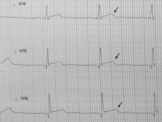
EKG showed changes suggestive of transmural lesion.
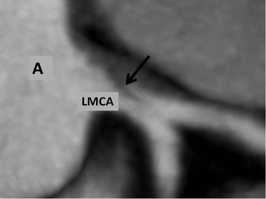
Multidetector coronary angio-tomography reveals dissection at the ostium of the left main coronary artery. A, aorta; LMCA, left main coronary artery; arrow, dissection.
The patient underwent emergent coronary revascularization with saphenous vein grafts to the first obtuse marginal and the left anterior descending artery (LAD). His postoperative course was uneventful and the patient was discharged home 7 days later. Control CT coronary angiography was performed 2 months later revealing patent aorto-coronary grafts (Fig. 3).
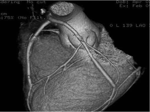
CT coronary angiography reconstruction showing patent grafts to the LAD and obtuse marginal arteries.
DISCUSSION
In 1931 Harold Pretty described the first case of spontaneous dissection of a right coronary artery during the autopsy of a woman who presented with precordial chest pain [1].
This condition is a rare cause of ischemic heart disease and affects mainly young healthy women. In 1987, 85 cases were reported in the literature, currently more than 300 cases have been published [2, 3].
Before the coronariography era, these cases were reported during autopsies of patients having sudden cardiac deaths and its incidence may have been under-estimated [2]. With the advent of these studies, the incidence has been reported to range between 0.07 and 1.1%. The LAD is affected in 60% of cases [3, 4]. Dissection of the LAD artery is most common in women, whereas in men it is the right coronary artery [2, 4]. The left main coronary artery involvement, like in our case, is rare, occurring in up to 12% of the cases [3], but is the most severe injury and presents as MI [5].
One-third of cases occur in women during the peripartum period. Hemodynamic and hormonal factors determine morphological changes in the arterial wall, and these may contribute to spontaneous dissection of the coronary arteries, with a peak incidence during the second week after birth [3, 4].
To date, the pathophysiology is unclear [2–4]. In a subgroup of patients it is not possible to identify a specific condition causing spontaneous coronary dissection and is therefore classified as idiopathic. In patients with connective tissue disorders such as Marfan’s and Ehlers–Danlos’ syndromes, predisposing to the dissection arises from medial degeneration of the coronary arteries [3].
Certain vasculitis, including systemic lupus erythematosus and polyarteritis nodosa, has been associated with the occurrence of coronary artery dissection. Other factors that can cause vascular spasm and coronary dissection include intense exercise, sneezing and prolonged cocaine abuse [2, 3].
Urgent coronary angiography is indicated if coronary dissection is suspected. It should show the presence of a double radiopaque lumen separated by a radiolucent intimal flap or a slow clearance of contrast from the false lumen [6].
However, coronary dissection may elude diagnosis by angiography. An intimal tear is present in only a minority of cases and a medial hematoma may not be recognized on coronary angiography as the medial hemorrhage may cause luminal narrowing or occlusion by pushing the inner media against the opposing wall [7].
Several cases in the literature have shown that CT coronary angiography provides additional information over invasive coronary angiography, with an accurate demonstration of the intimal flap and extent of the intramural hematoma. This study constitutes an emerging noninvasive alternative for diagnosis and also the follow-up of coronary artery spontaneous dissection [7].
There are no specific guidelines for the management of spontaneous dissections of the coronary arteries [3, 5]. Coronary angioplasty should be considered when the dissection is well located. Coronary artery bypass surgery is indicated in patients with multivessel dissection, failed angioplasty and dissection of the trunk of the left main coronary artery [3–5].
Although spontaneous dissection of a coronary artery is an uncommon condition, treatment should be based on the clinical status and imaging studies, including conservative and surgical means depending on the clinical presentation, location and characteristics of the dissection, we elected to proceed with surgery based on the CT findings.
ACKNOWLEDGEMENT
Special thanks to Jonh C. Mason for his assistance in the preparation of this manuscript.


