-
PDF
- Split View
-
Views
-
Cite
Cite
Marilyn Ng, Ruben Toribio, Gainosuke Sugiyama, Rare case of concurrent intussusception and volvulus after Roux-en-Y gastric bypass for morbid obesity, Journal of Surgical Case Reports, Volume 2013, Issue 1, January 2013, rjs018, https://doi.org/10.1093/jscr/rjs018
Close - Share Icon Share
Abstract
Gastric bypass patients are at risk for small-bowel obstruction secondary to adhesions, internal hernias, intussusception and volvulus. Most gastric bypass patients do not present with classic obstructive symptoms. We present a rare case of concurrent intussusception and volvulus in a woman with previous history of internal hernia following laparoscopic Roux-en-Y gastric bypass surgery.
INTRODUCTION
Small-bowel obstruction (SBO) is a common complication after bariatric surgery. Causes of SBO include adhesion, internal hernia, volvulus and intussusception [1]. Given the alteration of Gastrointestinal anatomy after bariatric surgery, diagnosis of SBO may be delayed and obscured by vague symptoms. We report a case of SBO presenting with concurrent intussusception and volvulus in a bariatric surgery patient. It is essential to have a high degree of suspicion to diagnosis SBO early and intervene promptly.
CASE REPORT
A 43-year-old woman [body mass index (BMI) 27.4 kg/m2] with a past medical history of morbid obesity (BMI 53.1 kg/m2) had progressively worsening upper abdominal pain and distention with associated bilious vomiting. She also reported constipation for 1 week.
Her past surgical history included a laparoscopic Roux-en-Y gastric gastrojejunostomy (LRYGB) and cholecystectomy 7 years ago. The bypass procedure was complicated 2 years later by SBO secondary to an internal hernia, which required exploratory laparotomy and repair. Four years after her LRYGB, a ventral hernia developed, which was repaired laparoscopically with mesh.
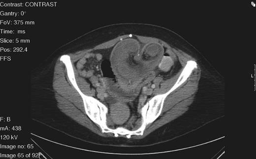
On examination, her abdomen was soft and distended with upper abdominal tenderness. No peritoneal signs were present. An upright abdominal X-ray demonstrated dilated loops of small bowel with air-fluid levels suggestive of SBO. Subsequent contrast-enhanced computed tomography (CT) of the abdomen and pelvis showed dilated loops of small bowel with evidence of strangulation and a mesenteric whirl sign converging at a suture line (Fig. 1).

Axial abdominal CT scan demonstrating a target sign mass consistent with intussusception with its inferior aspect tapering to a point consistent with volvulus.
An emergent laparotomy was performed. Intraoperative assessment revealed an infarcted volvulus segment of small bowel with twisted mesentery and proximal obstruction. The infarcted segment included the jejunojejunostomy suture line, which was resected and reconstructed. The surgical specimen demonstrated intussusception of small bowel with extensive mucosal necrosis. The patient had an unremarkable post-operative course and was discharged to home.
DISCUSSION
SBO is a recognized complication following LRYGB surgery for morbid obesity with a reported frequency of 0.2–4.5% [1]. Causes of SBO after bariatric surgery include adhesion, internal hernia, volvulus and intussusception. Unique to LRYGB is an increased rate of internal hernias compared with open RYGB [2]. Sites of internal hernias include the transverse mesocolon defect, Petersen's space and jejunal mesentery defect. Small-bowel volvulus has been observed in ∼17% of bypass patients [3]. Intussusception is a rare cause of SBO and represents ∼1% of all cases of SBO [4]. Intussusception is often a late complication and may be related to significant and rapid weight loss after RYGB [1].
Intussusception is either an anterograde or retrograde [5]. Retrograde intussusception is the most common type reported after both RYGB and LRYGB [5–8]. Anterograde and retrograde intussusception after RYGB may be caused by separate mechanisms, but the etiology of both remains unclear [7]. Others propose that a suture line might act as a lead point for intussusception [9], as we found in our anterograde intussusception. Another proposed cause of retrograde intussusception is disordered motility involving an ectopic pacemaker causing retrograde peristalsis [10]. In our patient, we noted a small-bowel volvulus and anterograde intussusception. To our knowledge, our case is the first concurrent presentation of intussusception and volvulus in an LRYGB patient presenting with SBO.
Both small-bowel volvulus and intussusception are often difficult to diagnose in the bariatric population. Patients will commonly present with abdominal pain and nausea. However, due to the small gastric pouch, voluminous emesis is rarely encountered [10]. The CT scan of the abdomen with oral contrast is the best diagnostic tool for patients presenting with SBO after RYGB [1, 5]. However, CT imaging may not readily differentiate between volvulus and internal hernia [10]. Nevertheless, a high index of suspicion is required to detect intussusception and small-bowel volvulus.
SBO is a common complication after LRYGB. Volvulus and intussusception after LRYGB for morbid obesity are both rare and challenging entities requiring a high index of suspicion, early diagnostic evaluation and surgical intervention. Due to the surgical alteration of Gastrointestinal anatomy following bariatric surgery, CT imaging should be obtained early to identify the potential cause of bowel obstruction. We report the first case of concurrent intussusception and volvulus in an LRYGB patient presenting with SBO.


