


Abstract
Meckel’s diverticulae can present with symptoms and signs mimicking acute appendicitis. We present the case of a healthy 35-year old male who was suspected of having acute appendicitis but at operation was found to have an inflamed Meckel’s diverticulum containing a giant faecalith, which had caused ischaemic necrosis due to a pressure effect. Post-operatively the patient admitted to a year of recurrent bouts of severe colicky abdominal pain, when running.
INTRODUCTION
It is well recognised that Meckel’s diverticulae can mimic the presentation of acute appendicitis and their presence must be sought in the case of discovery of an unexpectedly normal appendix at laparotomy. The presence of faecaliths within Meckel’s diverticulae are rare and seen in approximately 1% of all Meckel’s diverticulae. Giant faecaliths contained within Meckel’s diverticulae are extremely rare.
CASE REPORT
A 35-year old man presented with a 36-hour history of generalised abdominal pain, recently localised to the right iliac fossa. He had no significant past medical history and was taking no regular drugs. He had a low-grade pyrexia of 37.4 °C a pulse of 90 beats per minute, normal blood pressure and oxygen saturations and repeated clinical examination revealed increasing tenderness in the right iliac fossa with signs of localised peritonism. Blood tests demonstrated a mildly elevated white cell count of 12.6 x 10⁹ cells/L with a neutrophilia of 10.3 x 10⁹ cells/L and a CRP of 43 mg/L (normal level <10mg/L). The clinical picture was highly suggestive of acute appendicitis and the patient was taken to the operating theatre.
Free fluid was encountered on opening the peritoneum but the appendix was found to be grossly normal and after it’s removal a search was conducted for other pathology. A Meckel’s diverticulum was discovered approximately 30 cm proximal to the ileocaecal valve. It was dusky and thickened and contained a large hard mass. A diverticulectomy was performed using a linear stapler. The resected diverticulum was opened to reveal a large smooth faecalith 40mm in diameter. The histology confirmed a Meckel’s diverticulum with evidence of acute and chronic inflammation, haemorrhage and mucosal necrosis resulting in focal ulceration. The picture was that of an ischaemic type inflammatory pattern resulting from pressure from the contained faecalith.
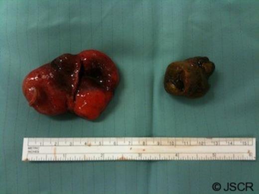
Resected Meckel’s diverticulum and the giant faecalith it contained
Post-operatively, the patient admitted that, for over a year he had suffered recurrent bouts of severe colicky central abdominal pain, when running. The patient made an uncomplicated recovery and was discharged home 2 days later.
DISCUSSION
Meckel’s diverticulum was described and named by Johann Friedrich Meckel, the Younger, (1781-1833), a Prussian professor of anatomy(1). It is the most common congenital anomaly of the gastro-intestinal tract and represents persistence of the vitello-intestinal duct, which is normally obliterated within the first 8 weeks of gestation. During early foetal life this duct connects the primitive gut to the yolk sac. It is a true diverticulum containing all layers of the intestinal wall. When present, it is situated on the ante-mesenteric border and may be connected to the umbilicus via a band.
Traditional teaching is that Meckel’s diverticulae follow the ‘rule of two’s’: i.e. they occur in 2% of the population, are two inches long and are located within 2 feet of the ileocaecal junction. Ectopic mucosa is encountered frequently and the most common type is ectopic gastric mucosa but ectopic pancreatic tissue can also be found (2). Carcinoid tumours are the most common malignancy encountered within a Meckel’s Diverticulum but adenocarcinoma and sarcomas also occur. The most common presentation in children is obstruction where the diverticulum forms the lead point of an intussusception. It can also present with rectal bleeding (due to ectopic gastric mucosal acid secretion causing ulceration to surrounding small bowel mucosa), or as diverticulitis mimicking appendicitis. If the Meckel’s diverticulum is connected to the umbilicus via a band, a volvulus can occur.
In adults the most common presentation is bleeding followed by obstruction and diverticulitis but the vast majority are discovered incidentally during operations for another indication (3).
Ninety percent of Meckel’s diverticulae occur within 90cm of the ileocaecal valve and the prevalence of Meckel’s diverticulae appears to be equal between the sexes but males seem more likely to develop symptoms with a ratio of 3:1 (4,5,6). A study by The Mayo clinic examined all Meckel’s diverticulae encountered over a 50-year period (n=1476). They found faecaliths in only 6% of symptomatic Meckel’s diverticulae and an even lower incidence in those that were asymptomatic. Overall they found 1% of Meckel’s diverticulae contained faecoliths equating to a total of 17 cases over 50 years. Acute (5,6) and chronic presentations (7) due to the presence of a faecolith within a Meckel’s diverticulum have been described in the literature.
While the case for removal of symptomatic diverticulae is clear (8), the question of whether or not to resect an incidentally discovered Meckel’s diverticulum remains debatable (9) with some advising against, especially in females (2). Some advocate removal of all Meckel’s diverticulae due to the low risk of post-operative complications versus the lifetime risk of developing problems with the diverticulum (10). Others suggest a selective approach with the decision to resect an asymptomatic diverticulum being positively influenced by a patient’s age (<50 years), male sex, diverticulum length greater than 2 cm or the presence of abnormal tissue within the diverticulum. Palpation of Meckel’s diverticulae is, however, reported to be unreliable in the detection of histologically abnormal tissue.
Acute presentations associated with Meckel’s diverticulae are a fairly rare phenomenon in the adult population and the presence of a giant faecalith within the diverticulum precipitating the presentation is extremely rare. The surgeon finding an unexpectedly normal appendix at operation must consider Meckel’s diverticulum and look for it. The decision to resect an incidental Meckel’s diverticulum is probably best made on a case-by-case basis, weighing up the risk of post-operative complications versus the lifetime risk associated with leaving the Meckel’s diverticulum in situ.
