


Abstract
Inguinal hernia is a common condition and may contain small or large bowel, omentum or other tissues. Leiomyomas of a round ligament are a rare condition occurring predominantly in premenopausal women. Abdominal, inguinal and vulvar locations have been described. Rare situations have been reported in which leiomyomas resemble to an incarcerated inguinal hernia. We describe a rare case of a leiomyoma of a round ligament in a young Caucasian female mimicking an incarcerated inguinal hernia. The treatment was surgical and we were able to remove the leiomyoma of the round ligament successfully. Leiomyoma of the round ligament is a benign tumor. Surgeons should take into consideration this condition in terms of differential diagnosis of masses that mimic an inguinal hernia.
INTRODUCTION
Leiomyoma of the female reproductive system is a common condition; however it is a rather rare tumor of the round ligament [1]. It usually occurs in premenopausal women, sometimes during pregnancy, and can be asymptomatic, or present with vague symptoms [2].
We describe a rare case of an incarcerated round ligament leiomyoma mimicking an inguinal hernia, diagnosed in a young female that was treated surgically, without the intraoperative presence of a hernia.
CASE REPORT
A 34-year-old Caucasian female presented to the surgical out-patient department of our center, complaining of swelling in the right inguinal region. She mentioned a gradual increase in size during the previous 3 months which was not reducible; however, no pain was present. Her medical history was free of other findings. Additionally, she denied any history of fever, weight loss or swelling in other parts of her body.
On examination, her vital signs and temperature were normal. Laboratory blood tests and radiologic imaging through abdominal X-ray also showed results within normal limits. Abdominal examination revealed a swollen, non-tender mass of dimensions of 6 cm × 6 cm in the right inguinal area. Attempted reduction of the mass was unsuccessful. Based on these clinical data, the patient underwent an operation in order to treat a possible incarcerated inguinal hernia.
Intraoperatively, we observed the presence of a round swelling arising within the superficial inguinal ring, whereas, while opening the inguinal canal, a mass that was attached to the round ligament was found (Fig. 1). The presence of a hernia sac was not identified, and the aforementioned mass was totally excised (Fig. 2). Pathologic examination of the postoperative specimen revealed the histologic characteristics of a leiomyoma of the round ligament. Postoperative period was uneventful and the patient was discharged the next day.
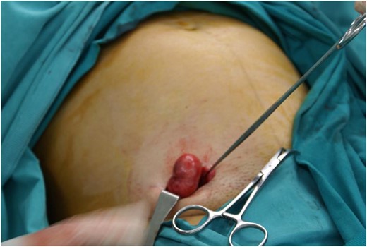
Leiomyoma of round ligament. Intraoperative finding.
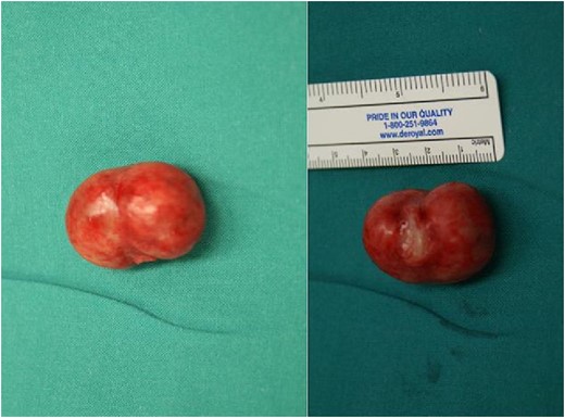
Excised round ligament leiomyoma. Postoperative specimen.
DISCUSSION
Leiomyomas are the most histologically common benign tumors of the round ligament, followed by endometriosis and mesothelial cysts [3]. They derive from smooth muscle tissues, can present as an inguinal or pelvic mass or other non-typical conditions [4], whereas their recent documentation is rare, possibly due to the rarity of the tumor [5].
An incarcerated leiomyoma of the round ligament presenting an inguinal hernia is an extremely rare situation; only few cases have been reported in literature [1]. Approximately, two-third of leiomyomas occurs in the extra-peritoneal portion of the round ligament and are more common on the right side for unknown reasons. A complex interaction between sex steroids (estrogen and progesterone) and local growth factors may interfere in the abnormal smooth tissue growth [3].
The broad differential diagnosis of inguinal swelling includes inguinal hernia, which is the most common diagnosis, but also tumors, cysts, abscesses, adenopathy and hydrocele of the canal of Nuck; surgeons should also take incarcerated round ligament leiomyomas into consideration, particularly in case of female patients. Preoperative imaging exams such as regional ultrasound, computed tomography (CT)-scan and magnetic resonance imaging (MRI) [6, 7] can help in diagnosis; leiomyomas appear as encapsulated heterogeneous masses on contrast CT and MRI images, associated with calcifications [8], but these findings are non-specific and do not lead to a certain preoperative diagnosis. In our case, further imaging investigation was not considered necessary preoperatively; however, the patient’s gynecologic history was free and did not mention the presence of other abnormalities, such as fibromas. Surgical excision is efficient for the treatment of the inguinal mass and proper diagnosis, followed by pathologic examination.
CONCLUSION
The presence of leiomyomas of the round ligament in situations that mimic irreducible inguinal hernia is a very rare condition. Surgical excision of the mass is considered the appropriate way of treatment. The accurate diagnosis is revealed intraoperatively and confirmed by histologic examination.
ACKNOWLEDGEMENTS
The authors certify that they have no affiliations with or involvement in any organization or entity with any financial or non-financial interest in the subject matter or materials discussed in this manuscript.
CONFLICT OF INTEREST STATEMENT
There has been no source of funding for the preparation of this manuscript. There are not any financial or non-financial competing interests.
