-
PDF
- Split View
-
Views
-
Cite
Cite
Ahmed M.A. Mohammed, Robert J. Dennis, Use of a venting PEG tube in the management of recurrent acute gastric dilatation associated with Prader-Willi syndrome, Journal of Surgical Case Reports, Volume 2016, Issue 1, January 2016, rjv174, https://doi.org/10.1093/jscr/rjv174
Close - Share Icon Share
Abstract
A patient with Prader-Willi Syndrome was admitted to the ICU with features of recurrent acute gastric dilatation, aspiration pneumonia and a massive pulmonary embolus. He was initially managed with intubation, assisted ventilation, intravenous fluids and anticoagulation. Decompression of the stomach was achieved with a nasogastric tube. After ventilator weaning, he did not tolerate the nasogastric intubation that led to a further episode of aspiration pneumonia as a result of non-resolving gastric dilatation. He required readmission to intensive care for a further period of ventilatory support. While the patient was sedated and ventilated, a venting percutaneous endoscopic gastrostomy (PEG) with a jejunal feeding extension was placed, permitting both continued decompression of the stomach and enteral feeding. The patient tolerated the PEG-J well and his nutritional needs were successfully addressed. Oral intake was slowly re-established with ongoing decompression of the stomach with the PEG. He was discharged from hospital with the PEG in place.
INTRODUCTION
Acute gastric dilatation is a severe complication of several pathological processes including but not limited to: abnormal eating patterns, small bowel obstruction, general anaesthetic and trauma.
It is a potentially lethal condition if not recognized early and managed promptly, as it can result in gastric ischaemia and perforation [1].
The treatment consists initially of gastric decompression and resuscitation, if this failed surgical management might become necessary, especially if peroration or ischaemia developed. Total gastrectomy is usually the preferred procedure as ischaemia of the remaining stomach is possible in cases of partial gastrectomy [2].
We present here a case of recurrent acute gastric dilatation that was treated successfully with a percutaneous endoscopic gastrostomy (PEG) tube during the acute presentation.
CASE REPORT
A 36-year-old-male patient had a background of Prader-Willi syndrome. He had the associated learning difficulties and hyperphagia seen with the syndrome. His dietary intake was carefully controlled within the care home where he lived and his body mass index was maintained at around 30. He also suffers from asthma and had orchidopexy as a child.
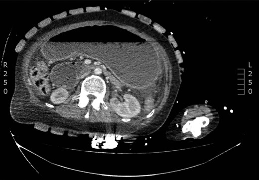
The patient was admitted as an emergency with abdominal pain and vomiting, following an episode of binge eating. A CT scan showed a grossly dilated stomach and a small volume of free gas in the abdomen (Fig. 1). Diagnostic laparoscopy did not reveal a perforation. His condition improved with antibiotics and decompression of the stomach with an NG tube. He was discharged after a 9-day admission.

The patient was admitted a second time, 7 months following the initial presentation with recurrent symptoms of abdominal pain, vomiting and respiratory compromise, after another episode of binge eating. The provisional diagnosis was of acute gastric dilatation with aspiration pneumonia and dehydration. He was admitted to the high dependency unit for stabilization, with oxygen, intravenous fluids and insertion of an NG tube was passed to decompress the stomach. The NG tube drained 1500 ml immediately. Fourteen hours after admission, he suffered a pulseless electrical activity cardiac arrest but was successfully resuscitated. A CT pulmonary angiogram and CT abdomen/pelvis showed a massive pulmonary embolus, pleural effusion, grossly distended stomach and ascites. The patient was sedated and ventilated in intensive care for 11 days. Four days after admission, a trial of NG feed was started. Despite the use of neostigmine and metoclopramide, NG aspirates remained high and total parenteral nutrition was subsequently started. He was discharged to the ward after 13 days in intensive care. Cautious oral intake was started after a swallowing assessment. He was unable to tolerate the NG tube and also found the central venous access distressing. After 16 days on the ward, he developed a further episode of respiratory failure and was readmitted to intensive care. The picture was of aspiration pneumonia and the patient required reintubation, ventilation and inotropic support. An upper gastrointestinal endoscopy showed significant residual food debris in the stomach, regurgitating to the oesophagus. The presumed aetiology of the recurrent aspiration pneumonia was gastric stasis causing food reflux into the oesophagus and bronchial aspiration.
After consultation with our local upper GI surgery unit, it was agreed that medium- to long-term decompression of the stomach would be required. As he was unable to tolerate an NG tube, a PEG tube with a jejunal extension was placed to vent the stomach and to permit enteral feeding.
The patient was successfully extubated 1 week later and discharged to the ward.
Over the course of a further 18 days, his oral intake was slowly increased with regular aspiration of the PEG-J tube.
He was subsequently discharged to the community, re-established on his normal dietary regimen and anitcoagulated with warfarin. At outpatient clinic follow–up, the PEG-J tube was planned to be removed under a direct vision at a further endoscopy.
DISCUSSION
Acute gastric dilatation is a well-described complication of the abnormal eating patterns associated with Prader-Willi syndrome. This can be difficult to diagnose as patients with Prader-Willi syndrome have high thresholds to pain and symptoms may only be evident at an advanced stage. The majority of case reports in the literature describe complications such as gastric necrosis necessitating gastrectomy [3]. We did not identify any previous case reports in the English literature, which describe the use of a PEG tube to decompress the stomach in this situation. Placement of the PEG tube in our patient proved to be critical in achieving effective decompression of the stomach over the short-to-medium time period. The risks vs. benefits of PEG placement must be carefully considered and while nasogastric decompression would remain our preferred route of gastric decompression, this case illustrates that in some patients, a PEG tube may be better tolerated and therefore a useful adjunct in the longer-term management of this challenging group of patients. We note that would be feasible for care home staff to learn how to manage episodes at an earlier stage in the community and perhaps stall the progression of such episodes, thus reducing the requirement for hospital admission for this vulnerable group.
CONFLICT OF INTEREST STATEMENT
None declared.
ACKNOWLEDGEMENTS
We thank Ms Jane Patterson, Consultant Upper GI Surgeon at Kings Mill Hospital, for sparing a valuable time for proofreading and providing invaluable advice on the case report. We also thank Toni Tuthil, the disability advisor at Peterborough City Hospital, who facilitated the communication with the patient.
REFERENCES
- anticoagulation
- pulmonary embolism
- aspiration pneumonia
- enteral nutrition
- intensive care
- intensive care unit
- intubation
- nutritional requirements
- patient discharge
- patient readmission
- prader-willi syndrome
- gastric dilatation
- ventilator weaning
- jejunum
- stomach
- dilatation of stomach, acute
- nasogastric tube
- nasogastric tube placement
- intravenous fluid
- percutaneous endoscopic gastrostomy
- gastrostomy tubes
- sedated state


