


Abstract
We present the case of a 32-year-old male with a delayed symptomatic left subclavian artery pseudoaneurysm secondary to protruding screws from prior perpendicular clavicular plate fixations. The pseudoaneurysm development and our operative approach are unique to the few similar cases available in the literature. The patient presented with a progressive pulsatile mass behind his left clavicle associated with paraesthesia. Angiography demonstrated a subclavian pseudoaneurysm adjacent to a prominent fixation screw. Offending screws were removed and the pseudoaneurysm was repaired using a reversed greater saphenous vein graft. This case illustrates an unusual aetiology for pseudoaneurysm development and highlights a successful operative intervention.
INTRODUCTION
Subclavian artery aneurysms are uncommon, yet the consequences may be life or limb-threatening [1]. True aneurysms, characteristically in later years, are typically degenerative. As opposed to pseudoaneurysms, which tend to be sequelae of vessel trauma. We present the case of a symptomatic pseudoaneurysm developing 9 years following multiple clavicular plate repairs for a non-healed clavicle fracture. A supra- and infraclavicular approach was used to remove offending screws and repair the pseudoaneurysm with an interposition vein graft. Our operative approach is unique to the few similar cases available in the literature.
CASE REPORT
A fit and well 32-year-old male was referred to vascular surgical outpatient clinic with a 1-year history of a progressive pulsatile mass behind his left clavicle, associated with intermittent paraesthesia. He had undergone plate fixation of a comminuted midshaft clavicle fracture 9 years ago. Due to non-union, the patient required a second plate inserted perpendicular to the first, with bone grafts for added strength.
On examination, there was an obvious pulsatile mass posterior to the left clavicle. The perpendicular plates were palpable, with prominent screws. At the time, all upper limb pulses were present with no signs of embolic disease. Neurological examination was also unremarkable. A recent Doppler ultrasound suggested that a plate fixation screw was impinging on the subclavian artery wall, causing increased wall thickness. Computer tomographic angiography (CTA) depicted a left subclavian artery pseudoaneurysm, yet there was significant metalware artefact. Thus, digital subtraction angiography (DSA) was booked (Figures 1 and 2). Yet, prior to undergoing this, he was admitted with sudden worsening of left arm pain and paraesthesia associated with movement despite a viable arm with present distal pulses.
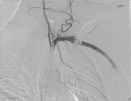
DSA oblique view illustrating fusiform left subclavian pseudoaneurysm.
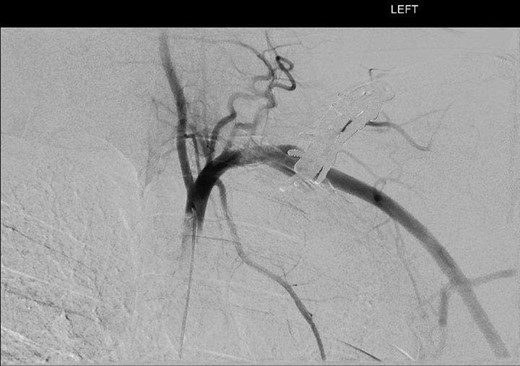
DSA illustrating fusiform left subclavian pseudoaneurysm.
Digital subtraction angiography via the right common femoral artery (CFA) was performed depicting fusiform aneurysmal dilatation of the mid left subclavian artery posterior to the clavicular plates and screws. Further, a focal area of irregularity was noted along the inferior aspect of the artery at the distal aspect of the aneurysm. On the dynamic study with arm in abduction there was significant compression of the artery by prominent clavicular screws at the site of the pseudoaneurysm.
The patient subsequently underwent open surgical repair of the pseudoaneurysm, with orthopaedic surgical assistance for removal of plate fixation screws. A supraclavicular incision was used for initial exposure. The clavicular head of sternocleidomastoid was divided and the phrenic nerve was distinguished and preserved after retraction of scalenus anterior. The subclavian artery pseudoaneurysm was identified posterior to the clavicular plates. There were two perpendicular screws from each plate protruding into the wall of the pseudoaneurysm. The proximal subclavian artery was looped and the clavicular plates exposed. At this stage, orthopaedic surgical assistance was required for removal of two screws from each plate. There was extensive bony overgrowth and the fracture was united. The plates were left in situ. An infraclavicular incision was then utilized to identify and loop the axillary artery before systemic heparinization. A reversed GSV interposition graft was secured end-to-end using 6/0 prolene to repair the pseudoaneurysm. The vein graft was tunnelled through the pseudoaneurysm. Further, the subclavian vein was explored to successfully exclude any damage to it.
The patient had a patent brachial pulse post procedure. Post-operatively the patient recovered well, without neurovascular impairment.
Upon review, the patient had complete resolution of symptoms and a full complement of peripheral pulses.
DISCUSSION
Subclavian pseudoaneurysms are a relatively uncommon phenomenon and typically occur as the result of blunt or penetrating trauma [2]. Invasive monitoring requirements utilizing central venous cannulation may inadvertently cause iatrogenic arterial injury leading to pseudoaneurysm formation [3]. Pseudoaneurysm development in our case presents the unique scenario whereby an existing true aneurysm caused by the initial injury enlarged over time to impinge upon the prominent clavicular screws. Iatrogenic vascular injury from plate fixation of clavicular fractures is rare, with only four similar cases available in the literature. Yet it appears all reports employed differing methods of repair.
Casselman et al. describes a subclavian pseudoaneurysm developing 8 years post internal fixation of a clavicular non-union [4]. The pseudoaneurysm was repaired with a prosthetic interposition graft, with exposure gained through a transclavicular approach including first rib resection.
Shackford et al. reported a case where a clavicular plate fixation screw had pierced the subclavian artery causing pseudoaneurysm development and brachial artery occlusion [5]. The patient underwent claviculectomy and interposition vein graft with autologous saphenous vein.
Similarly, Johnson published a case of left arm ischaemia secondary to a subclavian pseudoaneurysm where a compression plate screw was impacting the vessel [6]. Ligation of the subclavian artery coupled with carotid-axillary bypass was used to restore upper limb blood flow.
Albeit of the axillary rather than subclavian artery, Bain et al. also report a case of pseudoaneurysm development. This was also caused by arterial penetration from a plate fixation screw for a non-united clavicular fracture [7]. The patient underwent plate removal, then pseudoaneurysm repair via endovascular stent-graft with a good result.
Ranging from endovascular repair to open claviculectomy and first rib excision, all cases utilized somewhat differing surgical approaches. We initially considered using endovascular balloon occlusion to gain proximal and distal control of the subclavian artery, yet this idea was abandoned due to perceived obstruction of the operative field.
Due to the intrinsic neurovascular relationship of structures surrounding the subclavian artery, surgical intervention carries a high risk of neurological and vascular injuries [8]. As employed during our case, both supraclavicular and infraclavicular incisions provide adequate exposure to mobilize the proximal and distal subclavian artery. Due to the age and sporting pursuits of our patient, we managed to avoid more extensive bony resection such as claviculectomy, which would have granted greater exposure. If the proximal artery was involved, median sternotomy or thoracotomy may be required depending on the side [9, 10].
This is indeed quite a rare case that illustrates delayed iatrogenic arterial trauma. It is worth noting the importance of the clinical history and findings, both examination and investigations, in diagnosis. As found in the limited similar cases available in the literature, operative approach to this condition may vary and must be tailored to individual patient factors.
CONFLICT OF INTEREST STATEMENT
None declared.
