


Abstract
Sialolithiasis is one of the most common diseases of salivary glands in middle-aged patients. Sialoliths are localized in submandibular glands in nearly 80% of the reported cases and they are classified as ‘giant’ in case any dimension exceeds 15 mm. Giant sialolith in submandibular gland is a rare disorder. Here, an unusual case of giant sialolith in submandibular gland is reported. A 42-year-old man referred with complaints of recurrent pain and swelling in the left submandibular area. Computerized tomography revealed a calcified mass of 42 × 17 mm size within the submandibular gland. Excision was performed in the submandibular gland and a giant sialolith of 35 mm length localized in the body of the gland was detected. The postoperative period was uneventful and the patient fully recovered.
INTRODUCTION
Sialolithiasis is one of the most common diseases of salivary glands. Obstructing the salivary flow, sialoliths cause swelling, pain and recurrent infections of the associated gland. It is estimated that it affects 12 in 1000 of the adult population [1]. Sialolithiasis is more prevalent in middle ages and men are affected twice as many as women [2]. A greater proportion of sialoliths occur in the submandibular gland (80–95%), whereas 5 – 20% are seen in the parotid gland [3]. Intraglandular localization of salivary calculi is rarer when compared with intraductal localization [3]. Sialolith formation is more likely to occur in the submandibular gland due to its anatomic position requiring the salivary flow against gravity, longer and more tortuous duct and production of alkaline saliva rich in mucin. Most submandibular calculi are detected as radiopaque formations in plain radiographs and as radioluscent filling defects in sialography. Approximately 20% of sialoliths are not radiopaque, and sialography or sialendoscopy may be required to diagnose them. Salivary calculi are usually unilateral and do not cause dry mouth [2]. In the literature, sialoliths measuring >15 mm in any dimension or weighing 1 g are named as ‘giant’ [4]. Giant sialolith of the submandibular gland is rarely reported. A rare case of giant sialolith of submandibular gland, which is 35 mm in length, is presented in this report.
CASE REPORT
A 42-year-old male patient was admitted to our department with the complaints of recurrent pain, swelling and redness in the left submandibular area for 6 months. Swelling and hyperemia on the left submandibular region was observed. Intraoral bimanual palpation revealed a hard and tender mass, ∼6 × 5 cm in size. The orifice of Wharton duct was hyperemic and swollen, and the floor of the mouth was elevated in the left. A preliminary diagnosis of submandibular sialolithiasis was made and computerized tomography (CT) scan was performed. In the CT imaging, a calcified mass of 42 × 17 mm size in the left submandibular gland was detected (Fig. 1). Oral antibiotics and non-steroid anti-inflammatory drugs for 2 weeks were prescribed to treat the acute inflammation of the gland and a follow-up visit was planned. Two weeks later, it was seen that the acute inflammation of the gland had subsided and resection of submandibular gland was planned with the informed consent of the patient. Submandibular gland resection was performed. Intraoperatively, it was found that the body of the gland was totally filled with a giant sialolith and the gland parenchyma was atrophic (Fig. 2). Pathologic examination confirmed the diagnosis of chronic sialadenitis and a giant sialolith of 35 × 25 × 15 mm in size. The postoperative period was uneventful and the patient recovered without any complication.
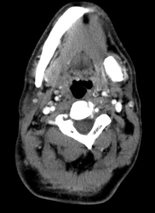
Coronal CT image showing the calcified mass of 42 × 17 mm in the left submandibular gland.
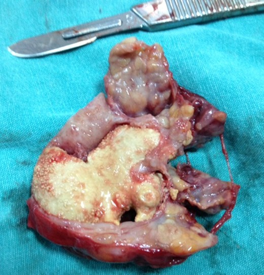
Intraoperative view of the giant sialolith located within the atrophic submandibular gland.
DISCUSSION
Although the exact etiology is unknown, sialoliths are thought to occur as a result of deposition of mineral salts around an initial nidus consisting of salivary mucin, bacteria or desquamated epithelial cells [2]. Stagnation of salivary flow, high alkalinity and increased calcium content predispose to sialolith formation. Salivary calculi are more often found in submandibular gland than parotid gland due to some factors such as the direction of salivary flow against gravity, a longer and more tortuous structure of Wharton duct and the higher calcium and mucin content of saliva produced in the submandibular gland [5].
Most calculi occur in Wharton duct, whereas the rest are found at the ductal hilum or within the gland [6]. Sialoliths of submandibular gland are rarely radioluscent and are recognized as filling defects in sialography. They were reported to be found more frequently in the left submandibular gland as in our case [7]. Sialoliths usually remain within the gland and enlarge but they rarely migrate to other locations. Sialoliths of submandibular gland detected in the oral cavity or fistulized to neck have been reported in the literature [8, 9].
Sialolithiasis of submandibular gland usually has an asymptomatic course. The symptoms begin whenever the lumen of Wharton duct is obstructed by a salivary calculus with resultant accumulation of saliva causing pain and swelling of the involved gland. The stasis of saliva can lead to bacterial ascent into the parenchyma of the gland and therefore recurrent infections. Long-term obstruction of salivary flow and recurrent infections can lead to atrophy of the gland with resultant loss of secretory function and ultimately fibrosis.
The primary objective of treatment for giant sialoliths is the restoration of normal salivary secretion. The treatment options for submandibular sialoliths are intraoral removal of stone, interventional sialendescopy and resection of the gland [10]. The appropriate treatment method is chosen according to the size, location and number of sialoliths. Small sialoliths can be removed through the duct orifice using bimanual palpation. Larger or proximally located sialoliths in the duct require surgical removal through oral cavity. Submandibular gland resection is indicated when a sialolith with a substantial mass (12 mm or more) is located within the gland and intraoral surgical access is not possible [10].
Newer treatment modalities such as endoscopic intracorporeal shockwave lithotripsy and extracorporeal shockwave lithotripsy have been introduced as alternatives to conventional submandibular gland surgery. Other techniques for sialolith fragmentation such as electrohydraulic and pneumoblastic devices have been described. Electrohydraulic devices have been criticized for the risk of injury to duct wall of the gland and there is no experience regarding the clinical use of pneumoblastic devices for the fragmentation of submandibular gland sialolithiasis. Sialendoscopy has been used as both a diagnostic and an interventional modality in salivary sialolithiasis. Small salivary intraductal calculi may be removed and biopsies may be taken using sialendoscopy systems. Submandibular sialoliths of such large sizes are extremely rare and should be treated after considering their size. Sialolithiasis can be managed by surgical or non-surgical therapeutic approaches. However, for giant sialoliths as presented in our case, submandibular gland resection remains the mainstay of treatment.
CONFLICT OF INTEREST STATEMENT
None declared.
REFERENCES
Author notes
This case report has been presented as a poster in 35th Turkish National Otorhinolaryngology, Head and Neck Surgery Congress organized in Maxx Royal Hotel, Antalya, Turkey, between 2 November 2013 and 6 November 2013.
