-
PDF
- Split View
-
Views
-
Cite
Cite
Nikolaos Arkoulis, Ben K. Chew, An unusual case of asymptomatic spontaneous umbilical endometriosis treated with skin-sparing excision, Journal of Surgical Case Reports, Volume 2015, Issue 3, March 2015, rjv017, https://doi.org/10.1093/jscr/rjv017
Close - Share Icon Share
Abstract
Spontaneous umbilical endometriosis is a rare extrapelvic manifestation of endometriosis. Very few such cases have been previously reported, almost always associated with a variety of symptoms, usually during menstruation. We present a case of asymptomatic umbilical endometriosis treated with skin-sparing excision. Differential diagnoses relevant to the clinician, as well as treatment options, are also presented. Surgeons should always consider umbilical endometriosis in their diagnostic approach when confronted with atypical umbilical nodules, regardless of whether they are symptomatic or not.
INTRODUCTION
Umbilical endometriosis, or Villar's nodule, is a rare, extrapelvic manifestation of endometriosis [1, 2]. Existing literature on this disorder reports a wide variety of symptoms associated with the umbilical lesions, ranging from mild to severe, usually cyclically synchronized with menstruation [1–7]. We present a case of completely asymptomatic umbilical endometriosis, adjacent to a previous peri-umbilical piercing site, which was treated by skin-sparing excision with a good postoperative result.
CASE REPORT
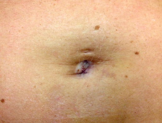
A 39-year-old female patient (para 1) was referred to the plastic surgery service from the department of dermatology with a nodular lesion arising from her umbilicus (Fig. 1). The lesion had been present for 18 months and had remained static in size for 1 year prior to presentation. It had remained asymptomatic throughout, with no episodes of bleeding, discharge or cyclical changes during menstruation. There was no history of previous trauma to the umbilicus with the exception of a long-standing peri-umbilical piercing. The patient had previously had excision of a benign ovarian cyst during caesarean section 10 years prior to presentation, but no other abdominal surgeries, in particular, no laparoscopic procedures with umbilical cannulation. She had not been diagnosed with endometriosis previously.

Preoperative appearance of the umbilical nodule. Note the previous piercing site at the 12 o'clock position.
Clinical examination revealed a firm polypoid lesion arising from the central umbilicus with a normal skin envelope. The lesion was in close proximity to the previous piercing but appeared to arise separately from it. Abdominal examination was otherwise normal with no clinical signs of hernia. Differential diagnoses at the time included keloid scarring, neurofibroma or dermatofibroma. Ultrasound scan of the lesion revealed a superficial soft tissue nodule, measuring 15 × 12 × 12 mm, with an identifiable posterior wall and no evidence of deep connections.
The patient underwent skin-sparing excision of the nodule under general anaesthetic. The resultant central umbilical soft tissue defect was reconstructed with a local flap fashioned from the skin-sparing excision. A good postoperative cosmetic outcome was achieved with no evidence of clinical recurrence 2 months after surgery (Fig. 2). Histology of the lesion revealed the presence of ectopic endometrial glands and stroma; hence, a diagnosis of umbilical endometriosis was made. She has since been referred to the department of obstetrics and gynaecology for further assessment.

Postoperative result 2 months following intralesional excision.
DISCUSSION
Endometriosis is a common disorder, defined as the extrauterine presence of endometrial glands and stroma. It is a common, oestrogen-dependent condition, affecting 5–10% of women in the reproductive age [8]. Endometriosis is associated with a wide range of symptoms, most commonly pelvic pain, dysmenorrhoea, dyspareunia and subfertility/infertility [5, 8]. Multiple hypotheses have been proposed for its aetiology, but its pathogenesis still remains unclear. Implantation of endometrial cells through retrograde menstruation, haematogenous or lymphatic dissemination of endometrial cells and the ectopic differentiation of pluripotent peritoneal progenitor cells to endometrial tissue (coelomic metaplasia) are amongst the most dominant theories of the pathogenesis of primary endometriosis [6, 8]. Secondary endometriosis as a result of the iatrogenic implantation of endometrial cells during surgery, especially cell seeding during laparoscopic port cannulation, has also been hypothesized [1, 7]. Endometriosis most commonly affects the pelvic cavity, but extrapelvic endometrial deposits affecting distant sites have also been widely reported [2]. Umbilical endometriosis, also known as Villar's nodule, is a rare extrapelvic manifestation of endometriosis, representing 0.5–1% of ectopic endometrial deposits [4, 7]. Few cases have been reported in literature, all associated with varying symptoms, most commonly size fluctuations during the menstrual cycle and discharge from the lesions. The presence of concurrent abdominal masses or previous history of endometriosis was also common in most cases, with imaging of the lesions, in almost all reported cases, showing intraperitoneal extension [1–4, 6, 7]. Extremely rare conditions such as recurrent catamenial pneumothorax have also been described [5].
In our case, the patient was a fit and healthy woman in her reproductive age with no previous symptoms or cyclical pain during menstruation. The presence of a previous peri-umbilical piercing further confounded the differential diagnosis, by adding keloid scarring or dermatofibroma to the list of possible pathologies. Ultrasound scan excluded intra-abdominal extension, though CT or MRI scanning would arguably have been more sensitive alternatives. Other disorders that need to be considered in the differential diagnosis of an umbilical nodule include malignancies (melanoma, sarcoma, adenocarcinoma, metastatic visceral carcinoma—Sister Mary Joseph nodule), inflammatory processes (abscess, omphalitis, folliculitis), other benign lesions (urachal anomalies, pyogenic granuloma, lipoma, haemangioma, inclusion cyst) and umbilical hernia [6, 7]. In terms of possible explanations of the pathogenesis of this extrapelvic deposit, the haematogenous or lymphatic dissemination of endometrial cells appears more likely than retrograde menstruation or coelomic metaplasia in this case.
Skin-sparing excision with local flap closure gave the patient a good postoperative outcome; however, in the event of recurrence, other surgical and/or medical modalities might need to be considered, such as complete resection of the umbilical stalk with or without surgical management of any pelvic/intra-abdominal disease and adjuvant hormonal therapies.
Atypical lesions arising in the umbilicus often pose diagnostic challenges. Surgeons should include endometriosis in their differential diagnosis in cases of atypical umbilical lesions and should bear in mind that cyclical symptoms are common but not always present. Simple skin-sparing excision and local flap reconstruction, when planned carefully, can give an aesthetically pleasing outcome, but contingency plans need to be in place in the event of recurrence.
CONFLICT OF INTEREST STATEMENT
None declared.


