


Abstract
A foreign body in the rectum is not a very common surgical emergency case. In the treatment of rectal foreign bodies, the aim is to use the simplest possible method while protecting the integrity of the intestine. Many removal techniques have been described in the literature. Here we report a case in which a transanal technique using a single incision laparoscopic surgery port was successfully used.
INTRODUCTION
A foreign body in the rectum is always a challenging diagnostic and management dilemma that begins with the initial evaluation in the emergency department and continues through the postextraction period. Usually, it is a result of pathological sexual activities performed by the patient themselves, or by other people, the latter being a criminal act [1]; however, sometimes, a foreign body is swallowed passes through the gastrointestinal tract and is held up in the rectum [2]. Most of them are excreted (80–90%), some require endoscopic removal (10–20%), but 1% require surgical intervention [3]. One of the most common problems encountered in the management of rectal foreign bodies (RFB) is the delay in presentation, as many patients are embarrassed and reluctant to seek medical care [4]. Most of these patients present to the emergency room after efforts to remove the object at home that may lead to a perforation or significant bleeding from the rectum. Hence, a stepwise approach that includes diagnosis, removal and postextraction evaluation is essential [4]. A single incision laparoscopic surgery (SILS) port with a shape adapted to the anatomy of the anus was used to remove a rectal foreign body, for the first time, in Korea [5], our case was done almost in a similar fashion and as far as we know the first to be reported in France and the third case worldwide.
CASE REPORT
A 70-year-old male with the history of psychiatric disorders presented to our emergency department after insertion of a cap of a bibber (nursing baby bottle) into his rectum. He attempted to remove it by himself but did not succeed. Upon presentation, the patient was stable, abdomen soft no peritoneal signs and digital rectal examination (DRE) was done with failure even to palpate the foreign body with the tip of the finger. The patient was taken to the operating room under general anesthesia, underwent rectosigmoidoscopy that revealed no damage to the rectum and the cap to be impacted in the rectum at 12 cm from the anal verge (Fig. 1). Accordingly, a nonsurgical technique was attempted; manual and endoscopic attempts were not effective. So the patient was put in a lithotomy position; after manual dilatation of the anus, a SILS port was introduced into the anal canal (Fig. 2) and the rectum was extended by continuous CO2 insufflation up to 10 mm Hg. A zero degree 5-mm laparoscope was then introduced, and under direct vision, the foreign body was removed using two laparoscopic grasping forceps and gently extracted from the rectum (Fig. 3). A postextraction evaluation using the SILS port showed small superficial mucosa ulcerations with no serious damage to the wall of the rectum. Two days later a follow-up rectosigmoidoscope was done and was normal except for the superficial injuries that were seen before. The patient was then discharged to be followed by the psychiatrist.
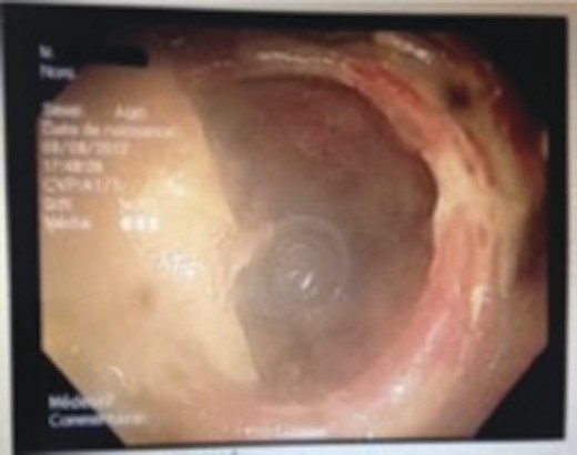
Aspect of the foreign body seen by endoscopy.
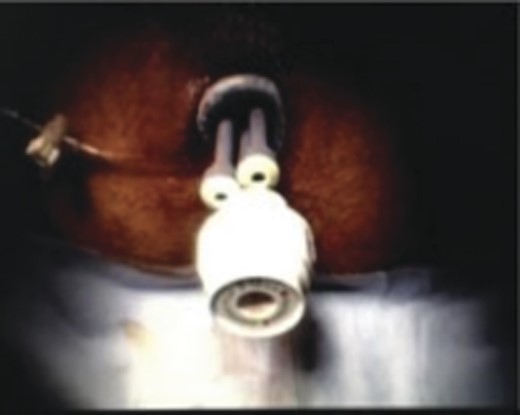
SILS port in the anus.
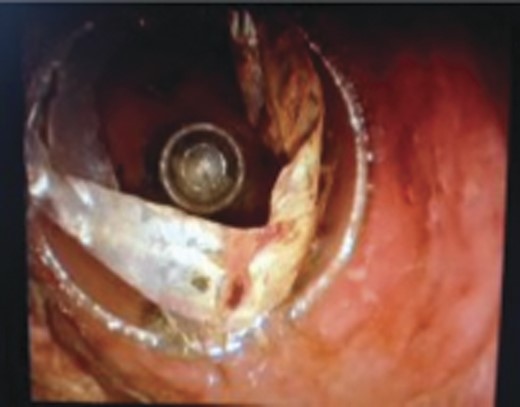
Foreign body seen by laparoscope.
DISCUSSION
RFBs present to the modern surgeon with a difficult management dilemma, as the type of object, host anatomy, time from insertion, associated injuries and amount of local contamination may vary widely. Management of these patients may be challenging, as presentation is usually delayed after multiple attempts at removal by the patients themselves have proved unsuccessful. Cohen and Sackier [4] showed a trend with 78% of cases linked to anal eroticism and 10% to assault. RFB may be high- or low lying depending on their location relative to the rectosigmoid junction. Generally, low-lying rectal foreign objects can be easily reached by the examining finger, whereas the ones above the rectosigmoid junction are difficult to reach. The distinction between high- or low lying retained objects is important for management. Generally, low-lying RFB that are palpable on DRE may be extracted in the emergency department. However, objects that are above the sacral curve and rectosigmoid junction are difficult to visualize and remove and may be unreachable even with rigid proctosigmoidoscopy. Fifty-five percentage of cases above the rectosigmoid junction require surgery, whereas only 24% required surgery when an object present in the rectum [6].
The basic principles of therapeutic intervention include transanal removal under the appropriate anesthesia, selection of patients for surgical retrieval, proctosigmoidoscopy following retrieval to assess the extent of damage and inpatient observation to rule out possible complications [4, 6, 7]. An anoscope or sigmoidoscope should be utilized to remove the foreign body under direct vision where possible to avoid iatrogenic injury [8]. There are varying techniques of transanal foreign body retrieval described in the literature including the use of polypectomy snares, forceps, inflated Foley balloon catheters, obstetric forceps, vacuum extractors, achalasia or dilation balloons and even plaster casts [9, 10].
A new approach to the management of high-lying rectosigmoid foreign bodies using the transanal SILS port technique with conventional laparoscopic instruments allows the direct vision and identifying the nature of foreign body, assessment of the rectal wall and the extent of injury if present. This approach is to avoid if intestinal perforation is suspected.
Another important aspect of this method is the replacement of the rectosigmoidoscope which is mandatory for postextraction evaluation of the distal bowel wall.
As a conclusion, in the treatment of RFBs, the aim is to use the simplest possible method while protecting the integrity of the intestine.
Outcome was excellent for this new method, which we think is simple and safe to use and can be considered as an alternative to surgical approach and even as a standard of care for removal of high-lying foreign body in order to avoid surgery.
