


Abstract
Filarial involvement of breast is a rare entity. Here is an unusual case of an old lady with right breast lump with axillary lymphadenopathy mimicking breast carcinoma, cytologically diagnosed as filarial mastitis. The patient subsequently received antihelmenthic therapy and showed marked clinical response within few weeks.
INTRODUCTION
Filariasis is an endemic disease in the Asian and African subcontinent and has emerged as a major health concern. It is caused by transmission of larvae of nematode Wuchereria bancrofti and Brugia malayi and it commonly affects lower limbs, spermatic cord, epididymis and retroperitoneum [1]. Here a rare case of breast filariasis has been reported.
CASE REPORT
A 50-year-old lady hailing from Bihar (India) presented to surgical outpatient department with the history of intermittent fever with chills for 6 months and gradually increasing right breast lump for 1 month. There was no history of nipple discharge. General physical examination was unremarkable. Breast examination revealed a large 8 × 8 cm breast lump involving all quadrants with well-defined margins, firm in consistency, bosselated surface, mobile (no fixity to pectoral muscle), the overlying skin was indurated, hyperpigmented with few dilated veins. Nipple was retracted on ipsilateral side and peau de orange was absent (Fig. 1). In the right axilla few pectoral group of lymph nodes was enlarged (2 × 3 cm). Rest of the systemic examination was normal. A provisional clinical diagnosis of inflammatory breast carcinoma was made.
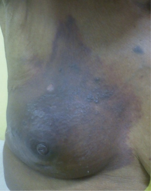
Right breast lump with diffuse pigmentation.
Fine needle aspiration cytology (FNAC) of the breast lump and the axillary lymph node was reported as a hemorrhagic aspirate showing many eosinophils, neutrophils and histiocytes along with microfilaria of W. bancrofti (Fig. 2), no malignant cells were seen and an impression of filarial mastitis was made. Differential leucocyte count was P69 L19 M04 E08 and the absolute eosinophil count was 1200 cells/mm3 (0–450 cells/mm3) corroborating with the cytology report.
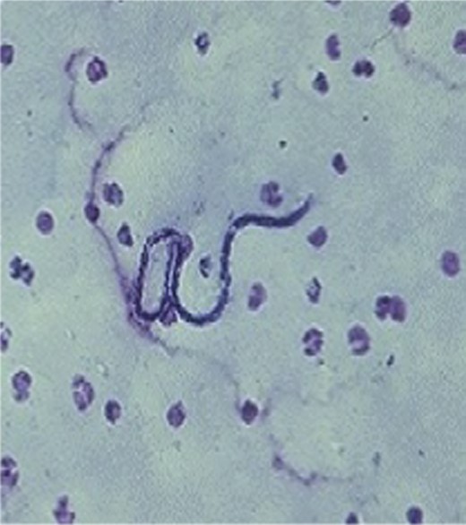
FNAC showing W. bancrofti against a hemorrhagic background.
Midnight peripheral blood smear showed—normocytic normochromic red blood cells, no microfilaria were seen. Ultrasound of the breast showed multiple cystic lesions in 3, 7 and 11 o'clock positions. There was no evidence of adult filarial worms. A final diagnosis of breast filariasis was made and the patient was started on diethyl carbamazine 6 mg/kg/day for 21 days. Following which the patient showed dramatic response with the resolution of the overlying pigmentation and a gradual decrease in lump size.
DISCUSSION
Filariasis is an endemic disease in India particularly Bihar (17%), Kerela (15.7%) and Uttar Pradesh (14.6%) [2]. Man is the definitive host of this parasite with a predilection for lymphatics. Culex mosquitoes serve as an intermediate vector when they feed from an infected person they ingest the microfilaria. These larvae develop into active motile forms penetrating the stomach wall and finally reaching the proboscis (10–12 days) ready for further transmission to a new host. The larvae finally develop into adult worms in the lymphatic system. The female worm gives rise to 50 000 microfilaria/day [3].
Some of the unusual sites of presentation of filariasis are breast [4], thyroid [5], body fluids [6, 7] and skin [8]. Walter et al. [9] suggested that microfilaria appear in tissue fluids and exfoliated surface material due to lymphatic or vascular obstruction and subsequent extravasation. This explains the possible mechanism of breast involvement in filariasis.
Our patient is one of the few rare cases which have been reported till date from India. The absence of microfilariae in the peripheral blood in this case corroborates with the observation that in endemic areas filariasis can exist without microfilaremia [10]. This is an unusual case of breast flariasis which is clinically mimicking inflammatory breast carcinoma, thereby creating a diagnostic confusion.
Therefore, if any patient residing in an area endemic for filariasis presenting with initial history of fever followed by breast lump with overlying skin induration and pigmentation, one should always keep in mind the rare possibility of breast filariasis.
