-
PDF
- Split View
-
Views
-
Cite
Cite
J Sloane, O Aziz, T McCullough, M Carter, G Lloyd, Identification of a terminal ileum carcinoid tumour during bowel screening colonoscopy – should terminal ileoscopy be performed as best practice?, Journal of Surgical Case Reports, Volume 2012, Issue 5, May 2012, Page 13, https://doi.org/10.1093/jscr/2012.5.13
Close - Share Icon Share
Abstract
The UK National Bowel Cancer Screening Programme invites men and women aged between 60 – 74 years old to be routinely screened every 2 years. A 90% caecal intubation rate or intubation of the terminal ileum is considered to be the best practice means of identifying completeness. This case report describes how terminal ileal intubation carried out during a routine screening colonoscopy led to the identification and treatment of a carcinoid tumour. Despite evidence for improving colonic diagnoses, completion of colonoscopy by passing through the ileocaecal valve is not performed routinely due to the perceived difficulty of the manoeuvre. With practice, ileoscopy has been shown to be achievable in at least 85% of routine colonoscopies and contributes significantly to quality assurance and to the diagnostic yield.
INTRODUCTION
The UK National Bowel Cancer Screening Program invites men and women aged between 60 – 74 years old to be routinely screened every 2 years (1). This is based on the finding that population screening using biennial faecal occult blood testing has demonstrated a 16% reduction in mortality from colorectal cancer (2). A UK pilot study has successfully demonstrated this as a feasible and reliable method of screening (3). Screening endoscopists are required to pass an endoscopy ‘driving test’ before being allowed to screen and have their screening data rigorously evaluated. A 90% caecal intubation rate, verified by visualisation of the ileocaecal valve and appendix orifice, or intubation of the terminal ileum is considered to be the gold standard means of identifying completeness. Of these, terminal ileal intubation with ileoscopy has the additional benefit of determining whether the source of bleeding is the distal ileum. We present a case where terminal ileal intubation carried out during a screening colonoscopy led to the identification and treatment of a pathology that if missed, could have a significantly worsened prognosis.
CASE REPORT
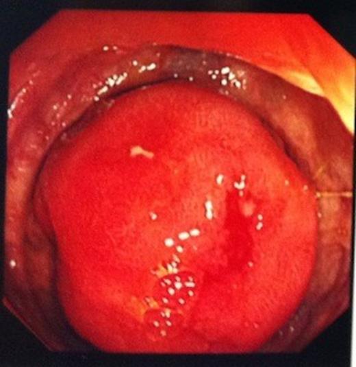
A 74 year old man was referred to our endoscopy unit after a positive faecal occult blood test. He had no preceeding symptoms or history that would place him at increased risk of colorectal cancer. At colonoscopy, excellent views were obtained to the ileocaecal valve (ICV) and appendix orifice, confirming identification of the caecum. With no colonic pathology seen, the endoscopist proceeded to intubate the ICV, as part of his routine practice. A 3cm ileal polyp was identified 5cm proximal to the valve (Figure 1) and biopsies were taken.

Histology demonstrated features of a classical neuroendocrine carcinoid tumour infiltrating the muscularis mucosa.

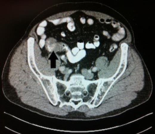
Abdominal CT scan, black arrow highlighting the carcinoid tumour in the distal ileum
CT scanning demonstrated the lesion intususscepting into the terminal ileum. There was no evidence of lymphadenopathy and no metastatic deposits were visualised. (Figure 2)

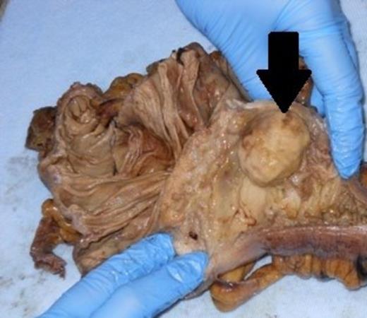
Resected specimen, black arrow demonstrating the carcinoid tumour in the opened distal ileum
The patient underwent a laparoscopic right hemicolectomy one month after diagnosis. The resected specimen demonstrated a carcinoid tumour measuring 32mm in diameter. Histological examination showed the tumour had breached the serosa into the pericolic fat. Twelve out of fifteen lymph nodes resected were involved with further evidence of extranodal spread. It was staged as PT3 N1 M0.
DISCUSSION
Carcinoids are neuroendocrine tumours derived from enterochromaffin cells most commonly found in the gastrointestinal tract (65% of cases) with an annual incidence of approximately two per 100,000 cases. Around 22% of cases present with distant metastases and in half these cases no primary tumour can be found (4).
Despite evidence for improving colonic diagnoses, completion of colonoscopy by passing through the ICV is not performed routinely. This is due to the perceived difficulty of intubating the valve as well as the anticipated increase in procedure time. With practice, ileoscopy has been shown to be achievable in at least 85% of routine colonoscopies. In skilled hands, it adds on average just additional 3 minutes to the procedure and contributes significantly to quality assurance and diagnostic yield (5).


