-
PDF
- Split View
-
Views
-
Cite
Cite
Kary Suen, Shiran Wijeratne, John Patrikios, An unusual case of bilateral verrucous carcinoma of the foot (epithelioma cuniculatum), Journal of Surgical Case Reports, Volume 2012, Issue 12, December 2012, rjs020, https://doi.org/10.1093/jscr/rjs020
Close - Share Icon Share
Abstract
Verrucous carcinoma is a rare, well-differentiated variant of squamous cell carcinoma with a minimal incidence of metastasis. Conservative treatment and local excision have been utilized although with high recurrence rates. We present a unique case of a 44-year-old male with bilateral foot involvement with recurrences after local excision, leading to management with bilateral forefoot amputations.
INTRODUCTION
Verrucous carcinoma is a rare, well-differentiated variant of squamous cell carcinoma (SCC) with a warty appearance. It is slow growing, exophytic, low-grade and with minimal dysplasia. Although it has a negligible incidence of metastasis, it is known for its aggressive local invasion, compressing underlying soft tissue but lacking destructiveness [1].
Verrucous carcinoma has been described in three main sites: the oropharynx, genitalia and feet [2]. Epithelioma cuniculatum refers to verrucous carcinoma found almost exclusively in the foot and was first described in 1954 by Professor Ian Aird [3].
Historically, ‘epithelioma cuniculatum’ was given when the nature of this tumour was uncertain. ‘Epithelioma’ implies a tumour of the epithelium and ‘cuniculus’ is translated as rabbit burrow, referring to its resemblance to the crypts of the tumour.
Epithelioma cuniculatum is almost always unilateral and rarely involves multiple sites. Our case study is of interest because it is only the second case of bilateral feet involvement with multiple epithelioma cuniculata previously described in the English literature.
CASE REPORT
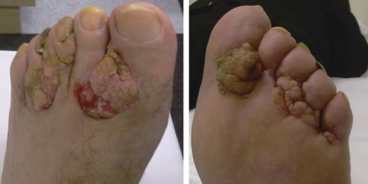
A 44-year-old male with a diagnosis of intellectual disability presents with a many year history of enlarging cauliflower-like tumours of both feet (Fig. 1).

Eight years prior to presentation, he had similar warty lesions resected from between the clefts of three toes on the left, with an amputation of one toe on the right. The histopathological report showed warty Bowen's disease with well-differentiated SCC.
The growths involved the interdigital folds of the toes of both feet. A CT scan showed tumour bony cortical disruption of the left second toe, with features suspicious for bone marrow invasion. The patient was further investigated with MRI, which showed tumour appearing to be closely related to bone and interphalangeal joints.
The extent of the lesions and possibility of bony involvement resulted in the decision to perform bilateral forefoot amputations.
The patient was followed up for 1 year and no further recurrences or metastasis were detected. He was able to ambulate with the assistance of orthotics.
Histologically, the specimen displayed both endophytic and exophytic morphology, comprising of warty papillomatous projections lined by variably atypical stratified squamous epithelium and overlying hyperkeratosis and parakeratin. The endophytic component showed deep acanthotic growths with a periphery of mitotically active basaloid cells and variable atypical squamous cells with frequent atypical mitoses. The endophytic downgrowths of tumour formed burrows filled with keratinous material, in keeping with verrucous carcinoma. No lymphovascular or perineural invasion was identified. Immunohistochemical staining for p16 showed strong nuclear and cytoplasmic reactivity, suggesting an human papillomavirus (HPV) related tumour.
DISCUSSION
The gross appearance of epithelioma cuniculatum is characteristic and distinctive, presenting as a warty, keratotic or sometimes soft tumour. Aird et al. described it as squashy, ‘with the consistency of an overripe orange’. A foul-smelling whitish sebum or toothpaste-like secretion can be expressed from the lesions [4, 5].
Most patients present in their 5–6th decades, with a predominance for men [6–8]. There is usually a delay in diagnosis due to the resemblance of the lesion to a wart or corn, which grows progressively despite topical treatment [6, 7]. The median time to diagnosis was 13 years [4] in one study and 16 years in another case series [6].
Bony invasion of verrucous carcinoma is a rare occurrence. The correlation of nuclear atypia in histology and bony invasion is unknown, with disparity in the findings. In Coldiron et al. [2], bony invasion was not associated with nuclear atypia of tumour cells.
Verrucous caricinoma, while being locally aggressive, rarely metastasize. There are only five records of metastatic epithelioma cuniculatum in the literature, all with metastasis to lymph nodes and lung in one patient [4, 7].
Some authors postulated that true verrucous carcinoma does not metastasize and the documented cases of metastatic epithelioma may actually be SCC. They also highlighted the uncertainty as to the amount of cellular atypia present in verrucous carcinoma and suggested that increasing amounts of atypia may be more indicative of SCC [8].
Mulitple lesions have rarely been documented in the literature [7]. One case report, Kathuria et al. report multiple lesions on a patient—one local recurrence in a previous excision site and an additional adjacent site [5].
Only one case in the literature describes bilateral epithelioma cuniculatum and this patient was treated successfully with excision and selective toe amputation [9].
There is contention regarding the role of human pappilomavirus types 1–4, 6, 11, 16 and 18 in the pathogenesis of verrucous carcinoma. Despite some specimens displaying oncogene expression or altered p53 activity, many authors discovered no evidence of human papillomavirus (HPV) DNA [2, 8].
Trauma and long-term irritation have been implicated as weight-bearing areas that have been found to be more frequently affected than non-weight-bearing area [4, 7].
In our case, the presence of HPV-type 16 DNA was detected in the specimen. Also combined with the bilaterality of the lesions, we postulate that environmental factors, including HPV, are likely to be a factor in the aetiology of the tumours.
The current mainstay of treatment is a surgical excision. Attempts in electrodesiccation, cryotherapy and laser ablation often result in tumour recurrence. In the largest published case series, there was a 19% recurrence rate for lesions treated with local excision [7].
Moh's technique, which microscopically controlled dissection allows total tumour removal with maximum preservation of normal tissue structure, has been reported to be successfully implemented on patients with epithelioma cuniculatum.
Amputations are necessary when the tumour is too extensive or recur after multiple attempts of local excision [4, 10]. According to the literature, amputations were more likely to be performed when the tumour infiltrates the soft tissue between metatarsal heads or there is evidence of possible bony involvement on imaging. In these cases, it becomes impractical to perform local excision with good clearance margins.
Although being a rare diagnosis, awareness should be raised of epithelioma cuniculatum, due to the slow growing but locally aggressive nature of the tumour. Bony involvement and metastasis is highly unlikely with this diagnosis. Due to its high recurrence rate, treatment may require multiple attempts at local excision, toe amputation and as a last resort, below knee amputation.


