-
PDF
- Split View
-
Views
-
Cite
Cite
Christopher Månsson, Britt-Marie Karlson, An unusual presentation of a solid pseudopapillary pancreatic tumor, Journal of Surgical Case Reports, Volume 2012, Issue 12, December 2012, rjs022, https://doi.org/10.1093/jscr/rjs022
Close - Share Icon Share
Abstract
Solid pseudopapillary pancreatic tumor (SPPT) is a rare tumor that constitutes 1–2% of all pancreatic tumors and most of the patients are young females. SPPT has low malignancy potential and radical resection is associated with good results and a high survival rate, even in cases with large tumors: the 5-year survival rate is estimated as 95%. This paper describes an unusual presentation of an SPPT discovered after blunt trauma to the abdomen during a basketball game. Computed tomography revealed a large tumor in the pancreatic head and the patient was operated by pylorus-preseving pancreaticoduodenectomy. The histopathologic examination indicated an SPPT with R0-resection and after 4 years there were no signs of recurrence.
INTRODUCTION
The use of computed tomography (CT) after trauma can reveal more than traumatic injuries and sometimes diseases that require treatment are discovered. In this paper, a case of a young girl with a rare tumor identified on a trauma-CT is presented.
CASE REPORT
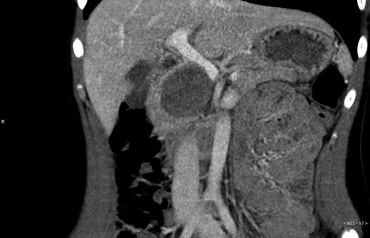
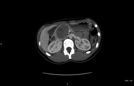
An otherwise healthy 14-year-old girl was referred to the Department of surgery Uppsala University Hospital from another hospital where she underwent a trauma-CT after receiving an elbow to the abdomen during a basketball game. The CT displayed no signs of traumatic injury, but revealed a 6.5 × 4.5 × 5.0 cm large tumor in the pancreatic head (Figs 1 and 2) which displaced the portal vein, vena cava and hepatic artery. Further investigation revealed that the patient had felt tired and had lost some weight during the past 6 months. Contrast-enhanced ultrasound with Doppler indicated a tumor with both solid and cystic structures, but no signs of vascular encapsulation. The investigation was complemented with positron emission tomography, hormone screening and an ultrasound of the heart. On the contrast-enhanced ultrasound the tumor appeared resectable; therefore no endoscopic ultrasound or biopsies were taken.


The patient underwent surgery, pylorus-preserving pancreaticoduodenectomy, and recovered well post-operatively and was discharged after 9 days. In the post-operative check-up 4 weeks after discharge, the patient was still doing well and histopathologic examination revealed an R0-resected solid pseudopapillary pancreatic tumor (SPPT).
The follow-ups were conducted at the referring hospital and MRI of the pancreas has been performed twice: 4 years after the operation, there were no signs of recurrence.
DISCUSSION
SPPT is a rare condition, constituting 1–2% of all pancreatic tumors. First described by Frantz [1], SPPT has been referred to by many different names, including Frantz tumor, papillary cystic neoplasm and solid and papillary epithelial neoplasm. The World Health Organization (WHO) has decided that the correct term is SPPT [2].
About 90% of SPPT patients are female, of which 85% are under the age of 35 years, with an average age of 24 years. Men generally tend to be older at the time of diagnosis [3]. As SPPT is predominant in women, studies have attempted to identify a connection between sex hormones and tumor genesis, but the results are inconclusive thus far [4]. As patients are often young, SPPT is suggested to be an embryonic tumor [5], although this has not been proved and further studies regarding its origin are needed.
SPPT has a low malignancy potential, but can turn malignant over time and the most common site of metastasis is the liver. Although the tumors are often relatively large, most can be R0-resected. In contrast to adenocarcinoma in the pancreas, the long-term results of SPPT are good, with an estimated overall 5-year survival rate of 95% [2, 6]. This motivates the use of surgery, even for large tumors. In a retrospective study, Campanile et al. [7] contacted all authors who had published reports about patients with incomplete resections between 1985 and 2008. In this small sample material, the median survival was 5.7 years [7] and the study highlights the need for R0-resection.
The pathology of SPPT is well described in a review article by Papavramidis and Papavramidis [2]. The tumor is often large, but the gross appearance appears to vary, with cystic, solid and necrotic areas being present, and a fibrous pseudocapsule often surrounds the tumor, demarcating it from the pancreatic tissue. Under light microscopy, there is a combination of solid, pseudopapillary and hemorrhagic cyst structures and on immunohistochemical staining there is usually b-catenin activity in both the nuclei and the cytoplasm.
In contrast to adenocarcinoma, there does not appear to be mutations in p53, k-ras and p16 [6].
Radiologically, SPPT has known features. In CT, a well-defined heterogeneous tumor with both cystic and solid areas is observed, with the cystic areas being central and the soild areas being peripheral [8]. Calcification in the tumor has also been described [9].
It is rare for trauma-CT to reveal a pancreatic tumor in a 14-year-old girl and it is even more uncommon to find an SPPT on a trauma-CT.
Besides this case report, three other patients with SPPT have been operated: one of these patients had a pylorus-preserving pancreaticoduodenectomy and the other two had distal resections. Two of the patients were women in their teens and the other one was a 32-year-old man. All had relatively large tumors (with a mean size of 5.2 cm), but the tumors were still R0-resected. All four patients were followedup and asked to complete a standardized questionnaire: the response was 100%. One patient required reoperation due to stomach volvulus and one takes pancreatic enzymes. Generally the patients were satisfied with their surgery and felt well. The follow-up was approved by Uppsala Ethics committee.
Conflict of interest statement
None declared.


