-
PDF
- Split View
-
Views
-
Cite
Cite
DJ Reilly, G Kalogeropoulos, D Thiruchelvam, Torsion of the gallbladder, Journal of Surgical Case Reports, Volume 2011, Issue 3, March 2011, Page 5, https://doi.org/10.1093/jscr/2011.3.5
Close - Share Icon Share
Abstract
Torsion of the gallbladder is an uncommon condition that is rarely diagnosed pre-operatively. Here, we present the case of a 76 year old male who was found to have a complete torsion of the gallbladder, and was successfully treated with cholecystectomy.
INTRODUCTION
Torsion of the gallbladder is a rare condition. Since its initial description in 1898 (1), there have been at least 500 reported cases (2). Here, we present a case of gallbladder torsion in an elderly male.
CASE REPORT
A 76 year old man presented to the Emergency Department with a six hour history of sudden onset of upper abdominal pain and vomiting. The pain was localised to the right upper quadrant, and was associated with several episodes of vomiting. He had no symptoms of obstructive jaundice, and no constitutional symptoms.
His past history included ileal ulceration diagnosed on capsule endoscopy, as well as gastro-oesophageal reflux and osteoporosis. His surgical history was significant only for an appendicectomy.
On examination, he was afebrile and vital signs were within normal limits. Abdominal examination revealed right upper quadrant tenderness and voluntary guarding. Murphy’s sign was positive.
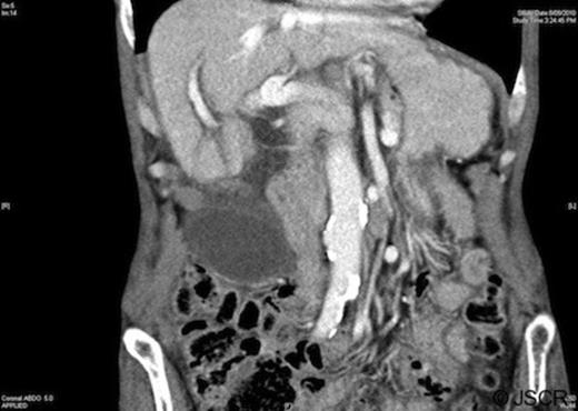
Blood tests were unremarkable, demonstrating a mild anaemia and normal liver function. White cell count (7.8×10⁹/L) and C-reactive protein (<5mg/L) were within the normal range. Abdominal ultrasound and computed tomography revealed a large distended, low-lying gallbladder with a diffusely thickened wall (up to 6mm), pericholecystic fluid and no cholelithiasis (Figure 1). The biliary tree was normal in calibre and ultrasound Murphy’s sign was positive.

Computed tomography demonstrating a low-lying gallbladder with a diffusely thickened wall and pericholecystic fluid
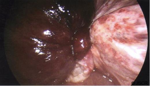
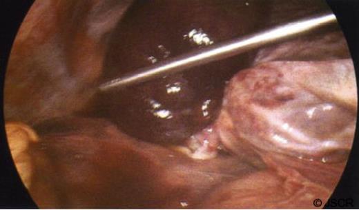
A diagnosis of acute acalculous cholecystitis was made, and the patient was referred for a cholecystectomy. At laparoscopy, he was found to have a gangrenous torted gallbladder (Figures 2 and 3), with a 360 degrees clockwise torsion. The gallbladder was detorted laparoscopically and an attempt was made at laparoscopic dissection, but Calot’s triangle was too inflamed and suffused with blood to clearly identify the anatomy and so the procedure was converted to open. The gallbladder was safely resected, and a cholangiogram revealed normal biliary anatomy with no bile duct stones.

Laparoscopic view of the gangrenous gallbladder, with a 360 degrees clockwise torsion on its mesentery

Laparoscopic view of the gangrenous gallbladder, with a 360 degrees clockwise torsion on its mesentery
Histopathology confirmed the diagnosis of gangrenous cholecystitis following torsion. The patient recovered well, and was discharged on day five postoperatively.
DISCUSSION
Torsion of the gallbladder remains a rare condition. The frequency has been observed to increase with age, with a peak incidence between 60 and 80 years (3). Women are more frequently affected (female to male ratio of 3:1) (4); although in children it is more common amongst boys (ratio of 1:4) (5).
The exact aetiology of gallbladder torsion remains unknown, although certain anatomical variants are thought to predispose to torsion (6). The first of these is where the gallbladder has its own mesentery; the second occurs when the cystic duct and artery have a mesentery, and the gallbladder is free within the peritoneal cavity. Given the age distribution of patients with gallbladder torsion, these congenital variants are thought to be exacerbated following liver atrophy and loss of visceral fat.
Pre-operative diagnosis of gallbladder torsion remains uncommon, owing to the rarity of the condition and the non-specific clinical and radiological features. Ultrasound will often demonstrate a thickened gallbladder wall with pericholecystic fluid. The gallbladder may appear to lie below its normal anatomic fossa, and may have an echogenic conical structure (representing the twisted pedicle) at the gallbladder neck (7). Stones may be an incidental finding (reported in 24.4% of cases in one study (8)), but are not thought to play any role in the aetiology of the condition. Similarly, computed tomographic scans may reveal an abnormal anatomical position for the gallbladder, as well as wall thickening and pericholecystic fluid.
Advances in imaging techniques, particularly multidetector computed tomography (MDCT) and magnetic resonance cholangiopancreatography (MRCP), have allowed an increasing number of cases to be diagnosed pre-operatively (2).
While rare, gallbladder torsion should be considered as a differential in elderly patients with right upper quadrant pain. A high index of suspicion, combined with improved radiological techniques, allows early diagnosis. With prompt surgical management, the condition has an excellent prognosis.


