-
PDF
- Split View
-
Views
-
Cite
Cite
L Lim, K Collier, R Harland, D Temperley, A rare cause of small bowel infarction, Journal of Surgical Case Reports, Volume 2011, Issue 11, November 2011, Page 9, https://doi.org/10.1093/jscr/2011.11.9
Close - Share Icon Share
Abstract
We report a rare case of small bowel infarction due to superior mesenteric artery occlusion secondary to cardiac tumour embolism. To our knowledge, this has not been previously reported in the literature. This case highlights a rare case and reviews current knowledge on the subject.
INTRODUCTION
Primary cardiac tumours are rare with incidence ranging from 0.001 % to 0.030 % (1). The most common malignancies affecting the heart are those due to secondary spread. This case highlights a primary malignant cardiac tumour with an uncommon complication (i.e. bowel infarction). Studies have illustrated cardiac tumour embolisation risk which varies from 12 % to 45 % (1). There have been no reports of cardiac tumour emboli resulting in mesenteric ischaemia.
CASE REPORT
A 53-year-old patient presented to hospital with a history of sudden onset excruciating abdominal pain in the umbilical region. The pain was sharp in nature and partly relieved by paracetamol and defecation. She also complained of abdominal swelling, nausea and vomiting. There was loss of appetite without weight loss. She had last opened her bowels one day prior to admission and had been passing flatus. She has reported to having had flu-like symptoms, chest tightness, cough and orthopnoea for one week prior to this admission.
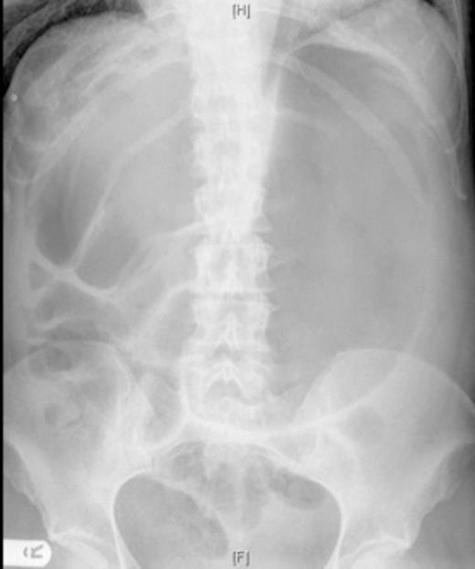
On examination, her abdomen was soft but distended, with tenderness in the epigastric region. The abdomen was hyperresonant on percussion. The patient had a chest x ray which demonstrated cardiomegaly and an abdominal x ray (figure 1).

AP abdominal radiograph; markedly distended stomach and distension of bowel loops in the right upper quadrant
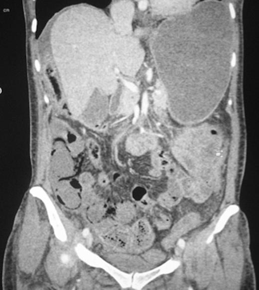
The patient subsequently underwent computed tomography (figure 2).

The patient underwent an urgent laparotomy within hours of admission. The small bowel was resected; a jejunostomy and mucus fistula were formed. It was found that the small bowel was non viable from about 80 cm distal to the duodenojejunal flexure to the mid ileum.
Second look laparotomy was performed after 48 hours. The bowel was judged to be non viable. A subtotal colectomy and revision of jejunostomy were carried out. Third look laparotomy was planned.
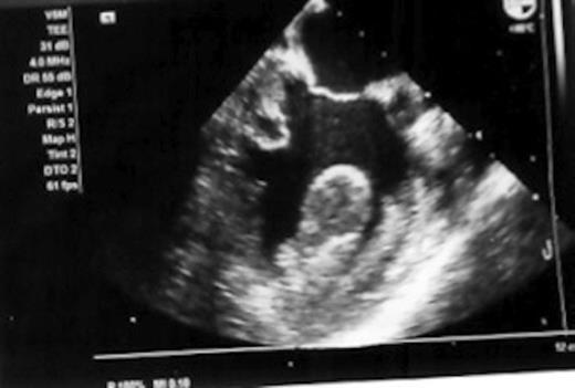
A postoperative transoesophageal echocardiogram (figure 3) was performed. This revealed impaired left ventricular function with a left ventricular clot. Specialist opinion on the echocardiography images was that the scans illustrated an extensive neoplasm in the left ventricle infiltrating both the myocardium and right ventricle leading to a diagnosis of sarcoma.

In view of the very poor prognosis, resection of the cardiac tumour was not advised. At this point, it was decided to commence supportive treatment. The patient passed away on the fifth day of admission.
DISCUSSION
Cardiac sarcomas account for 10% of cardiac tumours. Reardon et al (2) found that the majority of these tumours occurred in patients in the 40-50 age range. Prognosis is poor with median survival reported as 16.5 and 9.5 months in 2 studies which looked at 24 and 17 cases respectively (3,4).
Characteristics which are common with a malignant cardiac tumour were detailed by Silverberg (5) (i.e. Sarcoma) These are as follows: (i) right side cardiac mass is more likely to be malignant than left sided; (ii) Mass originating from free wall of cardiac chamber as opposed to the septum; (iii) Invasion of pericardium, great vessels and mediastinum; (iv)Presence of distant metastases; (v) extension of the mass into more than one cardiac chamber; (vi)concomitant pericardial and /or pleural effusion; (vii)diameter of mass more than 5 cm; (viii) tissue inhomogeneity and Contrast enhancement. These features ware present in the case we present with the exemptions being points iii and iv.
Elbardissi et al (6) noted that of those patients who suffered embolic events, 15% were associated with malignant tumours. It was noted that the tumour types most associated with embolism were aortic valve tumours and papillary fibroelastomas.
Acute mesenteric ischemia is an uncommon condition with a high mortality rate quoted to be between 60-90% (7).
Primary vascular causes can be equally divided between thrombotic, embolic and non occlusive. Embolic causes (as was the case with this patient) are commonly associated with atrial fibrillation where most thrombi arise from the left atrial appendage (8). The patient in this case was not found to be in atrial fibrillation.
The superior mesenteric artery is most commonly affected by emboli due to its acute angle of origin from the abdominal aorta. An embolus would characteristically lodge distal to the origin of the middle colic artery (9).
There has thus far been no documentation of mesenteric ischaemia/infarction occurring as a result of embolisation from a cardiac tumour. ElBardissi et al (6) reported 80 embolic events in their cohort of patients however not one patient was recorded as having an embolic event affecting the bowel.
Due to the significant embolic event and subsequent complications, the patient was not considered for resection of the cardiac tumour. Reshma et al (10) looked at 4 cases of patients with malignant cardiac tumours who underwent resection. Three of these were angiosarcomas and one was a high grade spindle cell sarcoma. All patients underwent post-operative chemotherapy. The outcome was poor in all these cases which are in contrast to the resection of benign cardiac masses.


