


Abstract
Small bowel obstruction (SBO) is a very rare complication post-caesarean section (CS). Herniation of small bowel through the rectus muscle with an intact sheath is extremely rare. We present a case of SBO after an uncomplicated c-section and an uneventful early postoperative course.
INTRODUCTION
The incidence of SBO after CS is very low (0.1%) (1). SBO secondary to herniation of segment of small bowel between intact rectus sheath and rectus muscle is extremely rare. This phenomenon, for which only two case reports exist in the published literature (2,3), could be related to non-closure of the parietal peritoneum. Despite the fact that non-closure of the parietal peritoneum at CS has become standard practice (4), many older obstetricians continue to advocate peritoneal closure as it has traditionally been associated with low morbidity. Here we present a case of severe post-operative morbidity that could potentially be related to non-closure of the parietal peritoneum at CS.
CASE REPORT
A 34 year-old caucasian woman (BMI 24.6) in her first pregnancy underwent an elective CS at 39 weeks gestation for breech presentation. Her antenatal course was uneventful. Surgery was performed under spinal analgesia via a lower transverse incision and a male infant weighing 3.15kg was delivered. Routine closure of the uterus and abdomen was performed, the parietal peritoneum was not closed. The patient’s short-term post-operative recovery was uncomplicated and she was discharged day 4 on simple analgesia.
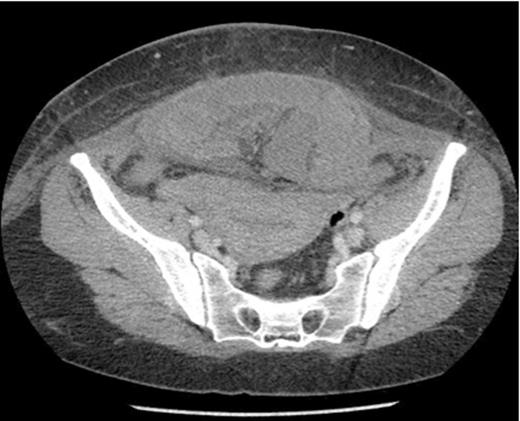
Axial section showing dilated loops of small bowel and a segment of small bowel adherent to the anterior abdominal wall.
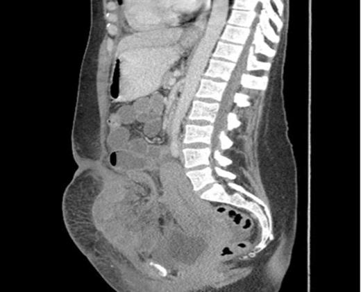
Sagittal section showing dilated loops of small bowel and a segment of small bowel adherent to the anterior abdominal wall.
On day 8 she presented to the Accident and Emergency Department with a five-hour history of severe lower abdominal pain with associated nausea, vomiting and one episode of diarrhoea. She was afebrile and physical examination revealed generalized abdominal tenderness and a well-healed wound. The uterus was tender and palpated at 24 weeks size. Vaginal examination was unremarkable. Lab results showed a neutrophilic leukocytosis (13) and mildly elevated CRP of 14.8. The initial working diagnosis was endometritis and the patient was admitted and commenced IV antibiotics (amoxicillin plus clavulanic acid and metronidazole).
The next day she became pyrexial (38°C) and tachycardic (103 bpm). Examination revealed a tender abdomen, with guarding and absent bowel sounds. Her neutrophilic leukcytosis increased from 13 to 27 and CRP increased to 159. She was commenced on gentamicin and a CT abdomen/pelvis was ordered. Radiology reported a high-grade acute SBO secondary to herniation of segment of small bowel into anterior abdominal wall defect. A laparotomy was performed and revealed a segment of strangulated small bowel lying above the rectus muscles and trapped beneath the intact rectus sheath. 34cm of necrotic small bowel was resected and an end to end anastomosis was fashioned. The patient made a good post-operative recovery and was discharged 9 days later.
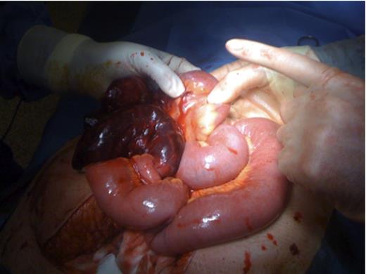
Operation pictures showing necrotic small bowel loop and dilated proximal small bowel.
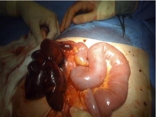
Operation pictures showing necrotic small bowel loop and dilated proximal small bowel.
DISCUSSION
The overall incidence of SBO after gynecological procedures is approximately 11% and 0.1% after cesarean section (1). The most common causes of post op SBO are adhesions and oedema. Internal herniation is an uncommon aetiology.
Current practice includes non-closure of the parietal peritoneum post CS. 66% of surgeons do not close the parietal peritoneum at CS according to national survey of practice in the UK (5). NICE recommendations suggest that neither the visceral nor parietal peritoneum should be sutured at CS. This is based on relatively weak evidence suggesting shorter operating time, less postoperative use of analgesics and improved maternal satisfaction (4). Shorter duration of the operation may have clinical benefits in terms of reduced risk of infection and postoperative complications such as paralytic ileus due to shorter exposure of the peritoneal cavity according to a recent Cochrane review (6).
However the recently reported CAESAR study, which was the largest randomized trial of caesarean section surgical techniques undertaken to date, demonstrate that no differences in short term febrile morbidity outcomes associated with closure versus nonclosure of the peritoneum at caesarean section (7). In addition a recent meta-analysis suggested that closure of the peritoneum was associated with decreased adhesion formation (8).
This case is an example of serious post-operative morbidity, which could potentially be avoided by peritoneal closure. Two similar case reports published previously (2,3) raise the question regarding appropriateness of non-closure of the peritoneum. Properly designed trials on long term morbidity would enhance the debate surrounding closure versus non-closure of parietal peritoneum post c-section.
REFERENCES
National Collaborating Centre for Women’s and Children’s Health Commissioned by the National Institute for Clinical Excellence. Caesarean section – Clinical Guideline April 2004
