


Abstract
Distal ventriculo-peritoneal shunt migration and extra-peritoneal CSF pseudocyst formation are unusual complications of shunt placement. We present a 65-year-old-female who received a subgaleal-peritoneal shunt to decompress a post-surgical subgaleal fluid collection. Eight weeks later, shunt series showed tight coiling of the distal catheter, and operative exploration found the distal shunt tip to have migrated superficial to the rectus sheath, where it had become encapsulated in a pre-peritoneal CSF pseudocyst. Migration of the distal catheter into the abdominal wall was likely due to local inflammation of the inner surface of the abdomen, with pressure from intestinal peristaltic movements and intra-abdominal pressure, and continued inflammation at the distal catheter tip may have caused formation of a pre-peritoneal CSF pseudocystic dilatation. To date, this is the first reported case of distal shunt migration into the abdominal wall with subsequent formation of an extra-peritoneal pseudocyst and represents a rare event in the surgical management of peritoneal shunts.
INTRODUCTION
The use of the peritoneum for cerebrospinal fluid (CSF) shunting was first introduced in 1905, and since then, ventriculo-peritoneal (VP) shunting has become a standard of treatment for hydrocephalus (1). Many complications of this procedure have been reported (2), with the most common complications being catheter obstruction, shunt infection, subcutaneous collection of CSF, peritoneal pseudocyst, bowel perforation, intestinal volvulus, mesenteric pseudotumor, migration of the catheter into the pleural cavity and heart, and several others (3). Abdominal wall migration and CSF pseudocyst formation are rare complications (4,5). In this report, we present a case of distal shunt migration into the abdominal wall at the left midabdomen with subsequent pre-peritoneal CSF pseudocyst formation. Possible mechanisms of this rare complication are discussed.
CASE REPORT
The patient is a 65-year-old-female with a history of recurrent left parietal hemangiopericytoma. One month after embolization and resection of the recurrent hemangiopericytoma, the patient presented with persistent fullness in the left mastoid area. This was significant for a postsurgical residual subgaleal fluid collection. There was concern that this might represent ongoing hydrocephalus. A subgaleal-peritoneal shunt with a Codman Programmable Valve was subsequently placed to decompress the collection and equalize pressure within the area. The subgaleal collection did resolve after shunt insertion. The peri-umbilical abdominal incision was closed in layers, and a tight purse-string was used to close the peritoneum around the distal catheter. Post-operative chest and abdominal radiographs confirmed placement of the shunt tubing over the left hemithorax and the distal shunt catheter within the peritoneum in the left lower quadrant (Fig. 1A).
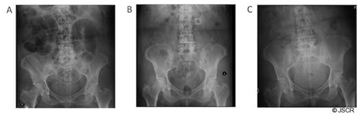
Distal shunt tip placement. (A) Abdominal radiograph demonstrates placement of the distal shunt tip within the peritoneum immediately post-operatively. (B) Distal shunt tip is shown tightly coiled and overlying the left midabdomen. (C) Distal shunt tip is again confirmed to be intra-peritoneal following distal shunt revision.
Two months later, the patient returned with complaints of headache and visual loss. CT scan of the head showed tumor recurrence in left parietal region, with recurrence of the subgaleal fluid collection, and shunt series showed the distal shunt catheter tightly coiled within the abdomen and overlying the left mid-abdomen (Fig. 1B and 2). The patient was afebrile, the abdomen was soft and non-tender with normal peristalsis and no signs of peritoneal irritation. Because of concern for hydrocephalus, the patient was taken back for distal shunt revision. However, due to the extent of tumor re-growth, the patient was not a candidate for tumor resection. Intra-operatively, the distal shunt catheter was found to have migrated superficial to the rectus sheath, where it had become encapsulated in a pre-peritoneal CSF pseudocyst. The distal portion of the tubing was cut off and new distal tubing was attached with a straight connector. The straight connector was subsequently stitched to the cyst wall to prevent future retraction. Abdominal X-ray reconfirmed peritoneal placement of the distal shunt tip (Fig. 1C).
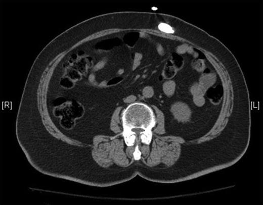
Pre-operative CT radiograph without contrast of the abdomen confirms extra-peritoneal migration of the distal shunt tip.
DISCUSSION
Intra-abdominal complications of subgaleal-peritoneal or ventriculoperitoneal (VP) shunting are uncommon. Complications have included: CSF pseudocyst formation, bowel obstruction, distal catheter extrusion, and perforation of viscera, all of which have been described previously. Distal catheter extrusion from the abdominal cavity have occurred through the umbilicus, rectum, the heart and pulmonary artery, intestine, uterus, vagina, abdominal wall scars, and other anatomical locations (2,5-8). A subgaleal-peritoneal shunt was placed for temporary decompression of the collection; however, in the case of hydrocephalus, a venticulo-peritoneal shunt would have been placed instead.
This patient presented with two rare abdominal complications with no apparent signs or symptoms of abdominal discomfort. The distal catheter had migrated from the peritoneal cavity to a location superficial to the rectus sheath and had become enveloped within a pre-peritoneal CSF pseudocyst. The mechanism of abdominal perforation likely includes erosion of the inner wall of the abdomen by apposition of the distal catheter against the abdominal wall from intrabdominal pressure, intestinal peristaltic movements, omental activity, and local inflammatory or foreign body reactions within the peritoneal cavity (2,5); however, the precise mechanism remains unknown. The type of catheter used may also increase the chance of this complication (5,7). For instance, extrusion of less flexible catheters, such as the Raimondi spring wire or simple silastic, have been reported in most cases, although extrusion of more flexible peritoneal catheters, such as the Holter and Pudenz, have also been implicated.
Pseudocyst formation is an unusual but well-described complication that is reported in <1% to 4.5% of VP shunts (9). A pseudocyst is composed of granulation and/or fibrous tissue that is not lined with epithelium and typically presents secondary to inflammation (3,10). Sheathing of the peritoneal catheter occurs subsequent to inflammation, and CSF drainage into the inflamed region ultimately produces a cystic dilation at the distal end. Pseudocysts have been reported to develop three weeks to five years after shunt placement, which is consistent with this patient’s presentation eight weeks after the shunting procedure. Treatment of this complication includes excision of the pseudocyst wall, removal and replacement of the distal shunt tubing, and possible re-direction of CSF drainage to another body cavity. Interestingly, previously reported CSF pseudocysts have been intra-peritoneal, while this patient presented with an extra-peritoneal pseudocyst. Previous reports suggest that migration of the distal catheter could be caused by local inflammation and necrosis of the inner surface of the abdomen, which in turn cause erosion and extrusion of the distal catheter into the abdominal wall. Intestinal peristaltic movements may then propel migration of the catheter, and continued inflammation at the distal catheter tip may cause formation of the pre-peritoneal CSF pseudocyst with subsequent cystic dilatation. However, in this patient, instead of necrosis of the abdominal wall, a more likely cause may be erosion of the peritoneal membrane around the (tight) purse-string with extra-peritoneal dislodgment of the catheter. Abdominal muscular wall sutural closure can open-up in the presence of excessive muscular contractions leading to further superficial migration of the catheter. The pseudocyst could be a proteinaceous seroma and may have the potential of being derived from tumor secreted fluid, especially since the shunt is a subgaleal-peritoneal shunt rather than a ventriculo-peritoneal shunt. While cytologic workup was not performed on the pseudocyst fluid, this should be strongly considered in similar cases. (3)
While there have been no reported migrations of a distal subgaleal-peritoneal or VP shunt to the abdominal wall with formation of a pseudocyst, such migrations are possible and within the spectrum of previously reported proximal migrations of the catheter.
