


Abstract
Total penile amputation is an exceptionally rare but devastating complication of circumcision, particularly when performed by untrained practitioners in resource-limited settings. We report the case of a 7-month-old male infant who presented 5 days after sustaining complete penile amputation during a traditional recircumcision. The initial procedure was conducted 1 month earlier, and the recircumcision was prompted by urinary difficulty, leading to complete penile loss at the penoscrotal junction. Examination revealed an exposed urethral stump without visible meatus. Due to delayed presentation and absence of viable tissue, replantation was not feasible. Urethroplasty with an 8 French nasogastric tube was performed, and the postoperative course was uneventful. The child resumed normal urination, with no complications at follow-up. This case highlights the serious risks of nonmedical circumcision in underserved areas and emphasizes the urgent need for regulation, community education, and access to trained surgical care and follow-up.
Introduction
Circumcision is one of the most frequently performed surgical procedures globally, carried out for religious, cultural, and medical reasons [1, 2]. While the complication rate is generally low, ranging from 1 in 500 to 2 in 100 cases, adverse events can range from minor issues to severe complications, including complete penile amputation [3, 4]. This case report presents a rare and devastating instance of total penile amputation in a 7-month-old infant following two traditional circumcision procedures. It underscores the critical risks associated with nonmedical circumcision practices and the challenges in obtaining timely and effective medical intervention in resource-limited settings.
Case presentation
A 7-month-old male infant presented with a 5-day history of penile amputation. One month prior, the infant had undergone an initial traditional circumcision. Five days before presenting to our facility, the caregivers observed that the child had difficulty urinating. Attributing the issue to an inadequate prior circumcision, they sought help from the same traditional healer, who then attempted a recircumcision. During this second procedure, a complete amputation of the penis occurred, resulting in moderate bleeding. There was no reported loss of consciousness; the bleeding was controlled at home before the child was brought to a healthcare facility.
Upon examination, the infant was alert with stable vital signs. A complete penile amputation at the penoscrotal junction was observed, with evidence of a healing wound and a previous repair site on the anterior aspect of the mid-scrotum. Urine was seen exiting through the exposed urethral stump, with no visible urethral meatus externally (Fig. 1).
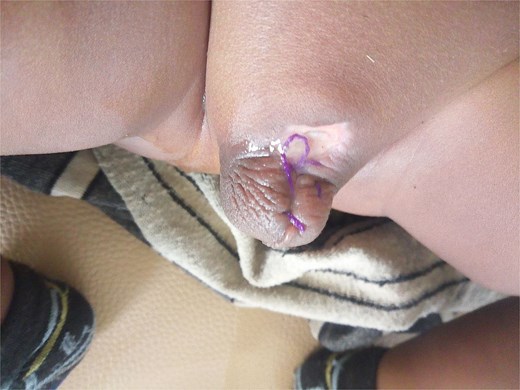
Total penile amputation with an anterior scrotal wound from attempted closure by a traditional practitioner; exposed urethral stump with no visible meatus.
Laboratory investigations, including complete blood count and renal function tests, were within normal limits. Urinalysis and urine culture were unremarkable. Abdominopelvic ultrasonography revealed normal kidneys and bladder, without signs of hydronephrosis or post-void residual urine.
The infant was admitted for supportive and surgical care. Intravenous ceftriaxone was administered to prevent secondary infection, and paracetamol was provided for pain management. The family was counselled about the severity of the injury and the potential need for advanced penile reconstructive surgery in the future. However, because of financial constraints, delayed presentation with nonviable amputated tissue, and the lack of microsurgical facilities in the region, a conservative management approach was pursued.
After obtaining informed and written consent, the infant underwent surgical exploration. The penis was found to be amputated at the penoscrotal junction. A previous repair site was noted on the anterior aspect of the mid-scrotum, but no external urethral orifice was identifiable. The previous closure was opened, and the proximal amputated urethral stump was circumferentially identified. An 8 French nasogastric tube (NGT) was inserted and secured as a urinary catheter, where it was left in place for 1 week to maintain urethral patency and facilitate the formation of a functional urinary tract through urethroplasty.
The postoperative period was uneventful, with no signs of infection, urinary retention, or other complications. Two weeks after surgery, the NGT was removed, and the infant urinated spontaneously without difficulty. At discharge, scheduled follow-ups were arranged. During the first follow-up visit, 2 weeks postdischarge, the infant continued to void normally, and the surgical wound had healed well without any signs of infection or stricture formation.
Discussion
Although circumcision is considered a relatively safe procedure, complication rates can increase significantly up to 20% when performed by untrained individuals [5]. A retrospective study conducted in Nigeria and Kenya reported that, among 50 hospitalized patients with postcircumcision complications between 1981 and 1998, 80% had been circumcised by untrained traditional surgeons. The outcomes included one death from septicemia, two cases of penile gangrene leading to total loss, and five cases of permanent disability from partial or complete glans or shaft amputation [6].
Penile injuries resulting from circumcision are classified into five grades:
Grade I: minor skin complications
Grade II: partial glans amputation
Grade III: urethral injury
Grade IV: penile fracture
Grade V: total penile ablation [7].
Our case represents a Grade V injury, the most severe form.
Common presentations of such injuries include acute hemorrhage, visible tissue loss, urinary retention, and pain. Most cases seek immediate medical attention, yet in this instance, presentation was delayed by 5 days. A comparable case documented by Suleiman Ayalew et al. involved a 46-day-old infant in Ethiopia who also presented 5 days postcircumcision with a complete penile amputation by a traditional healer [8]. The delay in presentation was likely due to caregiver misjudgment of the severity and systemic barriers to healthcare access.
Surgical options for penile amputation include:
primary re-anastomosis,
closure of the penile stump, and
Primary reanastomosis is feasible only when the amputated part is preserved and surgery is performed within 24 hours postinjury [9, 10]. In our case, the lack of tissue preservation and delayed presentation precluded replantation. Consequently, urethroplasty was the most appropriate step to restore urinary function, especially in settings with limited surgical resources [8].
Total penile amputation is a rare but profoundly distressing injury that presents surgical, psychological, and social challenges. Even in expert hands, management remains complex and carries significant long-term consequences for the patient and family [7]. The caregivers were counseled regarding long-term management strategies, including referral to a tertiary center with microsurgical capabilities for phallic reconstruction. Due to financial limitations, alternative plans were considered, such as delaying further interventions until the child reaches 4–7 years of age, when procedures like penile suspensory ligament release and phallic elongation may be performed. Ongoing follow-up is essential to monitor for complications such as urethral stenosis and to provide psychosocial support.
Conclusion
This case underscores the devastating consequences of circumcision performed by traditional circumcisers and the limitations imposed by delayed medical intervention. While urethroplasty restored urinary function in this infant, the long-term anatomical and psychosocial consequences remain unresolved. Preventive strategies, including community education, regulation of traditional circumcisers, integration into formal training programs, and improved access to safe surgical care, are vital to reducing the incidence of such preventable tragedies.
Conflict of interest statement
None declared.
Funding
None declared.
