-
PDF
- Split View
-
Views
-
Cite
Cite
Nadeem Chaudhry, Abid Qureshi, Mosammath Sultana, Meehika Pawanarkar, Muhammad Rizwan, A case of bone tumor-induced compartment syndrome, Journal of Surgical Case Reports, Volume 2024, Issue 7, July 2024, rjae470, https://doi.org/10.1093/jscr/rjae470
Close - Share Icon Share
Abstract
Compartment syndrome is a rare critical condition that can arise in individuals with cancer, presenting with significant challenges in diagnosis and management. Compartment syndrome occurs when the pressure within a closed fascial space rises to a point that restricts circulation. A 56 year-old male patient presented with 2 days of pain and swelling in the right upper extremity pain. Physical examination was remarkable for right upper extremity erythema swelling and tense compartments, concerning for compartment syndrome. Humerus X-ray showed moth eaten appearance of mid humerus with periosteal reaction and fracture. Patient was taken to the operating room for anterior and posterior compartment fasciotomies. Compartment syndrome is a surgical emergency, for which fasciotomy is generally performed. Pathology has rarely been linked to malignancy, with seldom reports examining causation. More research regarding pathophysiology of cancer in relation to compartment syndrome needs to be conducted.
Introduction
Compartment syndrome is a rare critical condition that can arise in individuals with cancer, presenting with significant challenges in diagnosis and management. Compartment syndrome occurs when the pressure within a closed fascial space rises to a point that restricts circulation [1]. Some pertinent causes of compartment syndrome are circumferential burns, long bone fractures, hematoma, and reperfusion of extremities that compromise tissue perfusion. Similarly, whether primary or metastatic, as bone cancer grows it can elevate pressure within the affected bone and tissue compartment, compromising blood flow and tissue health [2]. In cases of compartment syndrome in the setting of bone tumors, prompt recognition and interventions are vital to minimize tissue damage and enhance patient outcomes. Due to the intricate nature of treating compartment syndrome alongside bone cancer, a multidisciplinary approach involving oncologists, orthopedic surgeons, and other specialists is crucial for effective treatment and long-term care. Here, we present a case of compartment syndrome in the setting of a bone tumor.
Case presentation
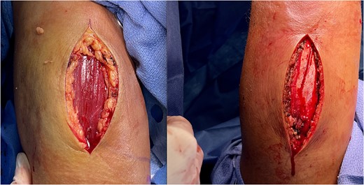
A 56 year-old male patient was evaluated at another facility for right upper extremity (RUE) pain after a fall. At the time, the patient underwent X-rays demonstrating lytic lesions associated with right arm humeral fracture. Patient was discharged to a subacute rehabilitation facility following conservative treatment. Over the last 2 days, the patient noted extensive pain and swelling so he was brought to the hospital for evaluation. Patient had a past medical history of Parkinson disease, hypertension, and lung mass of unknown etiology. Physical examination was remarkable for RUE erythema, associated with extensive swelling and tense compartments extending proximally from shoulder to elbow distally. Capillary refill was delayed with sensory deficits, pain was out of proportion to physical examination findings. RUE humerus X-ray showed moth eaten appearance of mid humerus with periosteal reaction and fracture, reflecting chronic osteomyelitis with pathologic fracture versus malignancy. Patient was taken to the operating room for anterior and posterior compartment fasciotomies of the RUE, following which the patient had recovered capillary refill time and sensation (Fig. 1). Postoperative course was unremarkable. Patient was offered biopsies of lung and bone during multiple hospital admissions, which were both refused.

Right upper extremity anterior compartment fasciotomy (left) and posterior compartment fasciotomy (right), with muscles exposed following release of fascia.
Discussion
Acute compartment syndrome (ACS), a critical limb-threatening condition, requires a prompt diagnosis and treatment. It relies on clinical assessment as well as pressure measurement. The associated clinical features have been classically defined as the 5Ps: pain out of proportion to physical exam findings, pallor, paresthesias, paralysis, and pulselessness [3]. Due to pain being a subjective entity, plausible cases can be false positives and make the diagnosis difficult to make. In the later stages, pain can significantly reduce due to paresthesia/anesthesia resulting from nerve ischemia and some rare cases of ACS present in the absence of pain [4]. The remaining four features are late signs after prolonged ischemia and subsequent significant neurovascular injury [5]. While clinical assessment has its place, quantitative investigations play a vital role in monitoring and promptly detecting ACS. The use of specific diagnostic tests, such as measuring compartment pressures using a handheld device called a manometer or performing intra-compartmental pressure monitoring, can confirm the diagnosis. The typical pressure within a compartment of healthy muscle is 10 mmHg of diastolic pressure and poses a risk when it approaches 30 mmHg and can cause permanent muscle vasculature and nerve damage due to compromised capillary perfusion.
There are several interpretations available for the pathophysiology of ACS. It can develop in regions of the body where there is limited ability for tissues to expand following trauma, muscle swelling, hematoma formation, and internal or external compression. The principal mechanism involves increased fluid pressure with intracellular and extracellular components. Subsequently, venous pressure rises, leading to a reduced arteriovenous pressure gradient, which decreases local blood flow [6]. After the leg, the most common location of ACS is within the forearm.
Rarely it can be a direct consequence of bone cancer and specifically sequelae of tumor growth. Bone tumors that grow rapidly or to a significant size can occupy space within the extremity compartment to cause local compression of adjacent soft tissues [7]. This includes, but is not limited to musculature, blood vessels, and nerves that further increase compartment pressure to compromise adequate perfusion. Ultimately ischemia can occur leading to tissue damage, further exacerbating compartment syndrome. It is important to note that bone tumor growth simultaneously stimulates an inflammatory response consisting of local swelling and edema. Finally, bone tumors can also cause venous obstruction, blunting venous return and similarly amplifying swelling and compartment pressure [8]. As such, early recognition and management of upper compartment syndrome in the setting of bone tumors is paramount to prevent irreparable tissue damage and widespread effects.
After a diagnosis of ACS, the implementation of treatment is time-critical. The primary treatment for ACS is fasciotomy; this relieves pressure and restores tissue perfusion. Immediate surgical fasciotomy is essential to prevent severe sequelae of the ACS. However, there was still controversy about the right time that fasciotomy should be done to avoid irreversible ischemic changes [3]. Unfortunately, fasciotomies are significantly associated with morbidities, including the need for further surgical management for delayed wound closure, surgical reconstruction with skin grafting or vascularized flaps, cosmetic problems, pain and nerve injury, permanent muscle weakness, and chronic venous insufficiency [4].
Prevention of compartment syndrome primarily involves reducing risk factors associated with the development of ACS [3]. It relies heavily on patient compliance with medical advice, particularly in terms of following post-operative care instructions or adhering to safe training practices in athletes. In athletes, proper training techniques, including gradual progression of intensity and duration of workouts, can help prevent overuse injuries that can lead to compartment syndrome. In the context of surgery, careful position and padding of the patient during prolonged procedures can help minimize the risk of developing compartment syndrome [9]. Monitoring and maintaining proper fluid balance and blood pressure during surgery are also important to prevent excessive swelling and pressure buildup in muscle compartments. Non-compliance significantly increases the risk of developing compartment syndrome or exacerbating an existing condition.
Literature surrounding compartment syndrome has been reported but not extensively studied. A case report of extranodal non-Hodgkin’s B-cell lymphoma was previously found in association with lower extremity ACS following a mechanical fall, for which fasciotomy needed to be performed [10]. One case–control study highlighted increased postoperative risks of ACS in populations of higher body mass indices and intraoperative serum lactate levels [11]. Marlborough and Venkataraman [12] have previously described a case of a patient with a hemorrhagic Baker’s cyst presenting with ACS, for which fasciotomy and debridement of abnormal tissue was performed, for which histopathology unexpectedly demonstrated soft tissue sarcoma. A limitation in our study was that the tumor diagnosis was never obtained but the induced pathological fracture alluded to malignant causes, which ultimately led to the increased compartment pressures and syndrome.
Conclusion
ACS is a surgical emergency, for which fasciotomy is generally performed to release compartment and lessen the pressures causing neurovascular entrapment. ACS has been linked to malignancy, with reports citing it as a risk factor and seldom reports examining causation. More research regarding pathophysiology of cancer in relation to ACS needs to be conducted, to examine patient populations more prone to the disease and preventive measures that can be undertaken to avoid morbidity.
Conflict of interest statement
None declared.
Funding
None declared.


