


Abstract
Although osteochondritis dissecans (OD) is well-described in the bibliography, cases of OD in the 1st metatarsophalangeal joint (MTP) are rare clinical situations. We present a 25-year-old male who presented to our department for big toe pain on the left side. Clinical and radiological examination showed the typical findings of OD lesion on the 1st metatarsal head (MH), establishing the diagnosis of OD. Intraoperatively the articular cartilage of the partially detached part of the MH was intact and under it, an empty cavity with a big defect was obvious. Autogenous cancellous bone transplantation from the metatarsal metaphysis, reattachment of the cartilage surface as a chondral flap and edge sealing with fibrin glue was our treatment of choice. OD of the 1st MH should be included in the differential diagnosis of big toe pain, as early diagnosis and treatment are crucial to prevent future osteoarthritis of the 1st MTP.
INTRODUCTION
Osteochondritis dissecans (OD) is considered to be a common cause of joint pain, not only among skeletally immature patients but in young adults too [1]. Although knee joint is the most typical presentation of OD, other joints can also be affected, such as elbow and ankle joint [2]. Treatment options include nonoperative and a wide variety of surgical methods, which depend mainly on the age of the patient, the clinical symptoms and the size of the osteochondral defect [3,4].
We describe a case report of a young adult (25 years old) with OD of the 1st metatarsal head (MH), treated with autologous cancellous bone transplantation and chondral flap coverage reattached with fibrin glue.
CASE PRESENTATION
A 20-year-old male presented to our department for progressive limping due to pain in the first metatarsal region (left). No history of trauma was present. Over the years, the pain was deteriorating till the level that even walking was severely impaired. In the last 6 months, the patient tried a nonoperative management including activity modification, shoe modification and orthotics. No relief of the symptoms was noticed.
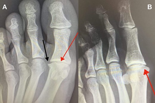
(A + B): (A) Anteroposterior, weight-bearing, X-ray view of the left foot. The red arrow shows a lytic lesion of the subchondral bone, indicating a possible OD lesion on the MH. The black arrow shows the formation of a small, lateral osteophyte. (B) Oblique X-ray view of the left foot. The red arrow shows again radiolucency of the cortical bone as well as small fragmentation of the MH.
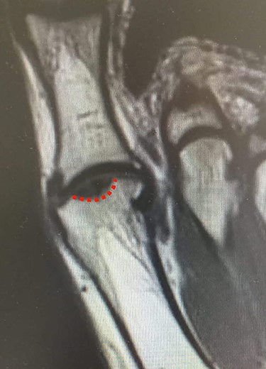
MRI of the left foot, shows low signal intensity lesion on the MH. The red dots show the depth of a subchondral cyst formation.
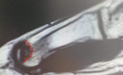
MRI (sagittal view) of the left foot. The red dots show a deep MH defect involving the subchondral bone, a typical finding of OD.
Clinical examination revealed localized pain on the dorsal side of the 1st MH and swelling of the 1st metatarsophalangeal joint (MTP). The range of motion of the 1st MTP joint was 40° of dorsiflexion and 30° of plantar flexion (slightly decreased). No pain was observed at the extremes of dorsi- and plantar-flexion. ‘Grind test’ was performed by compressing the 1st MTP joint and it was positive.
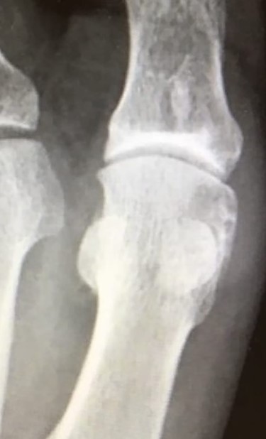
Standing anteroposterior (AP), oblique and lateral radiographs of the affected foot were taken. On the AP and oblique views, an OD lesion of the 1st MH was visible (lytic lesion of the subchondral bone and subchondral sclerosis formation) with a lateral-forming osteophyte (Fig. 1A and B). Magnetic resonance imaging (MRI) of the left foot clearly shows the osteochondral defect of the 1st MH (Figs 2 and 3).
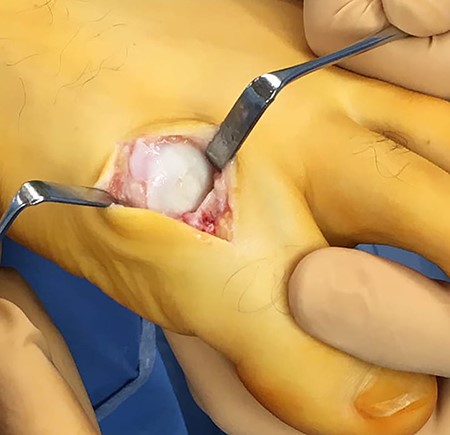
Operative treatment was our treatment of choice. A typical dorsomedial incision was performed to expose the 1st MTP joint of the left foot. Intraoperatively, a partially detached, nondisplaced from the surrounded bone cartilage, fragment was obvious (Stage-III classification of OD) [6]. Figure 4A shows the intraoperative finding of OD of the 1st MH. The detached bone was so loose that it could be separated from the surrounding MH very easily (Fig. 4B). The articular cartilage coverage was intact. Under the loose fragment, an empty cavity with a big subchondral bony defect and central depression into the subchondral bone was obvious (Fig. 4B). No signs of degenerative arthritis of the 1st MTP joint were obvious.
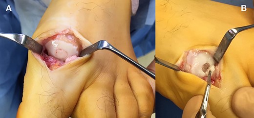
(A + B): (A) Intraoperative image, showing a central, partially detached, but not displaced from the surrounded bone OD lesion. (B) Under the OD fragment, an empty cavity with a defect and central depression into the subchondral bone was obvious.
After debridement and drilling of the lesion, autogenous cancellous bone from the metaphysis of the 1st metatarsal was taken to fill the empty cavity. Packaging of the void with autologous bone followed by reattaching the cartilaginous flap was performed. The edges of the cartilaginous flap were sealed with fibrin glue (Fig. 5).
Intraoperative image after filling the empty cavity with autologous cancellous bone. The edges of the cartilaginous surface were sealed with fibrin glue.
Postoperatively touch weight-bearing was allowed for 6 weeks with gradual full weight-bearing after 6 weeks. At 12 months follow-up, the range of motion of the 1st MTP joint was improved (50° of dorsiflexion and 30° of plantar flexion). An X-ray was taken, showing the restoration of the joint surface (Fig. 6).
Anteroposterior X-ray view of the left foot at 12 months follow-up showing a good-restoring articular cartilage and joint regularity.
Discussion
OD is a well-described pathologic condition in the bibliography that affects the subchondral bone and the overlying articular cartilage. It presents usually in skeletally immature patients with open physes (juvenile form) but cases of OD in adults are also reported [3]. Although the most common location of OD is the posterolateral aspect of the medial femoral condyle, OD lesions have also been found in the elbow (capitellum of the humerus), talus, wrist and femoral head [4].
Regarding the etiology of OD is still unclear, with acute trauma, repetitive microtrauma and local ischemia of the affected location being the main etiological factors [5]. The most commonly used classification of OD was described by Clanton and DeLee and classifies OD lesion in four Types, with Type I indicating a depressed osteochondral fracture, Type II a fragment still attached, Type III a detached but nondisplaced fragment and Type IV a displaced, loose fragment [6].
The treatment of OD of the 1st MH is the same as in other locations. The decision for operative management depends largely on four factors: the skeletal maturity, the size and the stability of the lesion and the clinical symptoms [4]. If nonoperative treatment fails then drilling and microfracture of the lesion to promote blood supply is indicated in the low-size lesions (<2 cm or Types I, II). In higher grade and size lesions (Type III, IV) osteochondral autograft transplantation, osteochondral allografting and autologous chondrocyte implantation are the best options [4].
Reporting cases of OD in the 1st MH are extremely rare [7–10]. According to Barlett DH., OD of the 1st MH not only can be painfully disabling but can also be the forerunner of adult hallux rigidus and suggests arthroscopic treatment [7]. Our patient, although did not have the typical signs of hallux rigidus, a formation of lateral osteophyte had already appeared. Camasta et al. [8] report a case of bilateral OD of the 1st MH, in a 43-year-old female and they state that if the articular surface is intact then packaging of the subchondral defect may be needed [8]. We also preserved the intact articular cartilage flap and reattached it with fibrin glue after debridement and subchondral bone grafting. Two more case reports with traumatic OD of the 1st MH are described in the literature that are treated with osteochondral autograft transfer [9,10]. Our cartilage flap technique with subchondral autogenous bone transplantation could be a less invasive treatment option.
CONCLUSION
OD of the 1st MH, although not common, should always be included in the differential diagnosis of big toe pain. It can cause pain and limited motion of the 1st MTP joint. A high index of suspicion, early diagnosis and early treatment are crucial because if OD lesions of the 1st MH are left untreated, can lead to degenerative, end-stage arthritis of the MTP joint.
CONFLICT OF INTEREST STATEMENT
None declared.
FUNDING
The authors received no financial support for the research, authorship and/or publication of this article.
INFORMED CONSENT
Informed consent was obtained from the patient for his anonymized information to be published in this article.
