


Abstract
A perforated retrocecal appendix resulting in a retroperitoneal abscess is a rare complication of a common disease. The first description of this condition was published in 1948. We present a case involving a 50-year-old woman who presented with abdominal pain inconsistent with the typical presentation of acute appendicitis and was eventually found to have a perforated retrocecal appendix accompanied by a retroperitoneal abscess. The patient was diagnosed using CT and operated upon but unfortunately had a resistant inflammatory process that led to persistent pus drainage from the abdomen despite multiple evacuation attempts and a prolonged hospital stay. In such cases, if the source of this type of inflammatory process has not yet been controlled or even identified, we recommend a second surgical examination, with additional surgical examinations as needed, and offer other suggestions.
INTRODUCTION
Appendicitis is a common cause of acute abdomen, with an expected lifetime occurrence of 7% and a mortality rate of less than 1% if the appendix is not perforated [1]. Despite recent technological advances, appendicitis continues to be diagnosed clinically [1]. The appendix is typically in the intraperitoneum, either anterior or retrocecal; however, in 30–65% of appendicitis cases, it may be hidden in a retroperitoneal location [1, 2], which could alter the clinical presentation, exacerbate challenges associated with diagnosis, particularly with late presentation, and increase complications [1, 3]. Thus, a retroperitoneal appendix can be described as ‘a snake in the grass.’ [4]
The first description of this condition was published in March 1948. [5] A subsequent article published in 1955 explicitly described the possibility of retroperitoneal appendicular rupture [6]. To date, around 30 case reports have been published, but the magnitude of this problem is still unknown [1–10].
Here, we report a case involving this condition, suggest an approach for this issue and offer recommendations to improve the understanding of this problem.
CASE DESCRIPTION
A 50-year-old woman presented to our hospital with a history of abdominal pain for 10 days that was mild and colicky in nature, mainly at the right flank and associated with nausea and vomiting.
After 10 days, the patient’s pain worsened, and she collapsed while walking. She was transferred to our hospital, with an acute surgical abdomen.
A CT scan revealed an appendix with an enhanced oedematous wall, adjacent retroperitoneal subhepatic air and mild collection indicative of a perforated subhepatic appendix (Fig. 1).
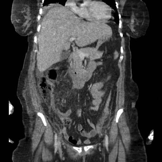
Coronal section CT scan image of the patient’s abdomen.
The patient was diagnosed as an acute abdomen due to a perforated appendix associated with peritonitis and sepsis.
The patient underwent an urgent laparotomy, which revealed pus that was evacuated. The appendix was found to be retrocecal, subhepatic and perforated.
Retrograde appendectomy was performed. Culture and sensitivity swabs were taken. The abdomen was washed extensively. Suction drains were secured to the right abdomen, and the surgery was not complicated.
The patient was admitted to the surgical intensive care unit, and she was managed accordingly, with an estimate of recovery and discharge within 10 days.
The patient was treated with antimicrobials, based on a culture and sensitivity report.
The patient did not improve as expected. In fact, her drains continued to drain 200 ml of pus daily despite appropriate coverage with antimicrobials.
Unfortunately, the patient developed respiratory distress, and was intubated and mechanically ventilated.
A second abdominal CT scan showed an ill-defined retroperitoneal collection, mainly on the right side. In fact, two pockets of collection in the right retroperitoneal area were observed: the first pocket was just under the abdominal muscles and peripherally, and the second pocket was just anterior to Gerota’s fascia of the right kidney.
An ultrasound-guided drainage procedure successfully drained 300 ml of pus.
The patient’s condition improved, and she was extubated and discharged from the ICU to the regular ward after a total ICU stay of 19 days.
While in the ward, the patient accidentally removed her drains; a repeated CT scan showed well-defined enhanced collections in the right and left retroperitoneal spaces with evidence of the formation of a fat-pus level indicative of infectious fat necrosis.
The patient was offered another drain attempt; she refused this procedure and chose to be discharged from our facility, where she spent a total of 36 days of hospital stay.
The patient was sent home on cefepime, vancomycin and metronidazole.
DISCUSSION
When the appendix is retrocaecally positioned, appendicitis is the primary suspect when the uncommon but potentially fatal complication of retroperitoneal abscess is observed [3].
Most patients with retroperitoneal abscess due to perforated retrocaecal appendix present insidiously. Pain is a significant presenting symptom in approximately 50% of cases, but none have complained of typical symptoms of acute appendicitis; instead, all of these patients have had unusual complaints [3, 7]. A retrocaecal retroperitoneal appendix may induce flank pain rather than the much more common symptom of right lower quadrant pain (RLQP) [1]. For an abscess resulting from a perforated appendix to reach the retroperitoneum, it must perforate through the parietal lining of the peritoneum [8].
With mortality of up to 16.7%, which is attributed to the development of peritonitis and sepsis, and an average hospital stay of 27.3 days [3], this condition must be better understood to achieve best practices.
Our patient showed signs of a persistent focus of infection that was not properly drained, either surgically or radiologically. Hsieh et al. [2] stated that ‘surgical management is mandatory but may or may not be effective.’ They recommended surgical management as the initial option because the patient is typically in more critical condition and because the appendix has to be removed; as the patient improves, radiological drainage might be attempted [2]. Based on our experience, a second surgical examination after all treatment modalities have failed might be mandatory and effective; thus, this approach might be the third option for patients whose conditions are refractory.
To support the concept of a second surgical examination, a patient had nonspecific abdominal symptoms for 32 years and underwent multiple interventions during that period eventually developed an appendico-cutaneous fistula that was diagnosed as a perforated retrocaecal retroperitoneal appendix [9].
The early use of MDCT scans in diagnosing complicated cases of appendicitis is important [9, 10]; such scans help to facilitate early management prior to the commencement of fatal complications; and they are far superior to ultrasound, conventional CT and even MRI when it comes to optimal abscess drainage approach [8].
RECOMMENDATIONS
In an unusual presentation and the absence of RLQP and tenderness, laboratory examinations to assess for leucocytosis, elevated C-reactive protein and neutrophilia can increase sensitivity to 97–100% [1].
An early MDCT scan, should be considered in cases involving suspected retrocecal appendicitis.
A second surgical examination, of refractory inflammatory processes needs to be part of the approach.
Effective communication between different specialists must occur to determine diagnosis, management plan and minimize complications.
ACKNOWLEDGEMENTS
None
CONFLICT OF INTEREST STATEMENT
None declared.
