-
PDF
- Split View
-
Views
-
Cite
Cite
Adam O’Connor, Peter Asaad, De Garengeot’s hernia with appendicitis—a rare cause of an acutely painful groin swelling, Journal of Surgical Case Reports, Volume 2019, Issue 5, May 2019, rjz142, https://doi.org/10.1093/jscr/rjz142
Close - Share Icon Share
Abstract
De Garengeot hernia is a rare subtype of femoral hernia whereby the vermiform appendix is located within the hernial sac. Even rarer is the presence of appendicitis within the hernia sac. De Garengeot’s hernia is difficult to diagnose pre-operatively and can prove technically difficult at operation particularly with regards to mobilization of the caecum and appendix in order to perform appendicectomy. Laparoscopic, open, with and without mesh repair of de Garengeot hernia have all been described in the literature with varying degrees of success. We present a case of an 82 year old lady presenting with an acutely painful right sided groin lump. CT scan revealed the presence of de Garengeot hernia with acute appendicitis. We describe in text and photo format our approach to the hernia repair, appendicectomy and provide a short review of the literature with regards to the different operative approaches to such a patient.
INTRODUCTION
De Garengeot hernias are a rare subtype of a femoral hernia which contains the vermiform appendix, accounting for 0.5–1% of all femoral hernias [1]. Even rarer is the occurrence of appendicitis within the De Garengot hernia diagnosed on pre-operative computed tomography scanning. Estimated incidence of acute appendicitis within de Garengeot hernia is 0.08–0.13% [1].To our knowledge, there are less than 100 case reports of De Garengeot hernia in the literature and only 3 of those with diagnostic CT findings prior to operation [2–4].
CASE REPORT
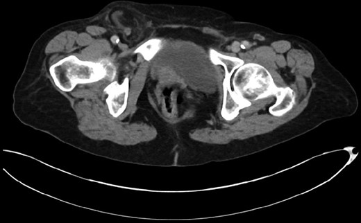
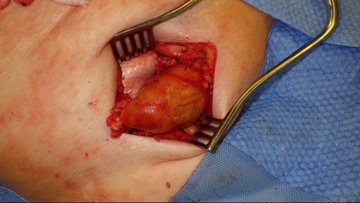
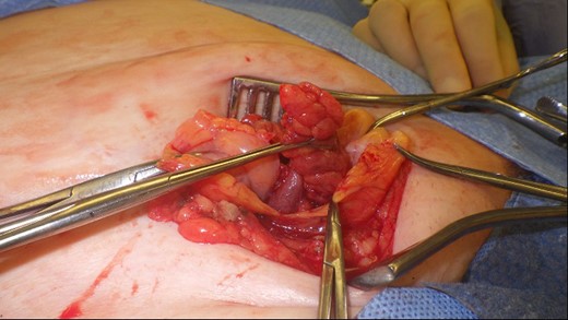
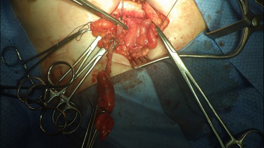
This 82-year-old lady presented with a sudden painful right sided groin swelling not previously noticed. She had no features of obstruction and was opening her bowels and not vomiting. Her medical background included left sided femoral hernia repair in 2005, coronary artery bypass graft and bilateral total knee replacements. On examination her abdomen was soft with the presence of a tender, irreducible swelling in the right groin, inferolateral to the pubic tubercle. She had good bowel sounds and there was stool present in the rectum on PR examination. A full set of blood tests demonstrated no abnormality. A CT abdomen and pelvis demonstrated an incarcerated right sided femoral hernia containing an 8 mm long inflamed appendix with a small amount of localized free fluid and inflammation indicative of De Garengeot’s hernia with underlying acute appendicitis (Fig. 1). The hernia sac diameter measured 2 mm on CT scan. She was taken to theatre for an open Lockwood repair of her femoral hernia and an appendicectomy. Following an initial Lockwood incision over the lump, dissection was performed down to the hernia sac also exposing the inguinal ligament (Fig. 2). The tightness of the femoral ring made mobilization of the appendix difficult. By partially incising the inguinal ligament superior to the femoral ring, the appendix was freed, and on inspection showed inflammation particularly towards the tip (Fig. 3). The caecum was then reduced and the inguinal ligament was repaired with non-absorbable suture. The femoral hernia was then repaired with a small funnel of ultrapro mesh. Appendicectomy was then performed in the usual fashion via the Lockwood incision leaving a slightly longer stump than usual (Fig. 4).

CT scan of the abdomen and pelvis demonstrating acute appendicitis within a femoral hernia.

De Garengeot’s hernia – femoral hernia sac containing the vermiform appendix.

Vermiform appendix freed from hernial sac showing inflammation towards the tip.

Excised appendix specimen with a slightly longer than usual appendix stump.
The patient had an uneventful recovery and was discharged on the second postoperative day. Subsequent histopathology of the appendix specimen demonstrated serosal inflammation throughout the appendix with partial fibrotic obliteration of the appendix lumen. No formal follow-up was organized and to date there have been no complications following surgery.
DISCUSSION
First described in 1731, the De Garengeot hernia is a highly rare subtype of the femoral hernia accounting for 0.5–1% of femoral hernias [1]. However acute appendicitis present in a De Garengeot hernia is even rarer with a case first described in 1785 and less than 100 cases reported presently [1]. As found in femoral hernias, the de Garengeot hernia is more common in women. The presence of a large and mobile caecum has been thought to pressure the appendix into the femoral canal thus causing de Garengeot hernia [2]. To our knowledge there only four reports of CT scan confirming the diagnosis prior to operation [3–5]. Often any peritonism due to underlying appendicitis is masked by the narrow hernia neck excluding the appendix outside the peritoneal cavity. Complications such as strangulation, perforation and abscess are all documented in the literature [6].
Given its rarity, there is no preferred surgical technique to the de Garengeot hernia. Watkins et al. reported performing an initial appendicectomy and interval mesh hernia repair and Voitk et al. reported an initial open drainage to drain an appendix abscess contained within a femoral hernia followed by interval appendicectomy and hernia repair [7, 8]. In both cases there were favourable clinical outcomes. Both laparoscopic and open approaches have been described with mesh both being and not being used. As described in our case, there is often difficulty mobilizing the appendix and confidently identifying the base which sometimes necessitates lower midline laparotomy in order to mobilize the base of the appendix.
Thomas et al. demonstrated the use of diagnostic laparoscopy as a method of assessing groin hernias which then demonstrated de Garengeot’s hernia [6]. Klipfel et al. also favoured laparoscopic appendicectomy in a de Garengeot hernia with an inaccessible caecum and long appendix before proceeding to Rives mesh repair of the femoral hernia in the same operation [9]. Given the paucity of the condition, no systematic reviews or RCTs have been performed to examine the best approach to take.
In our patient, once the appendix was freed from the sac we had numerous options. Consideration was given to reducing the freed appendix, repairing the femoral hernia and closing the initial incision before proceeding to a laparoscopic or an open appendicectomy. Given that we had reached the base of the appendix uneventfully, an open appendicectomy via the same incision was an equally safe yet more minimally invasive option for the patient. Additionally, we could have avoided repairing the femoral hernia in the emergency setting and just perform appendicectomy. This would have the benefit of allowing us to use mesh once the infection had settled but the disadvantage of recurrence preoperatively. Suture repair of the hernia alone would also have been possible but with a high chance of recurrence. Mesh repair of the hernia would have a significant change of infection. Given that the appendix was not perforated and there was no pus present, there was a very minimal risk of infection and that the risk of hernia recurrence was more pressing. We therefore decided to opt for a mesh repair of the hernia.
To our knowledge, this is one of only four cases demonstrating CT proven De Garengeot hernia with underlying appendicitis prior to operative intervention. The condition remains difficult to diagnose and can prove technically difficult in the operating theatre especially with regards to mobilization of the appendix and caecum. We demonstrate the benefit of incising the inguinal ligament in order to free up the caecum and appendix which in this case worked, allowing conventional appendicectomy to be performed.
CONFLICT OF INTEREST STATEMENT
None declared.
REFERENCES


