


Abstract
The left atrial appendage (LAA) is thought to be responsible for the vast majority of embolic strokes, and has become an important target in the surgical management of atrial fibrillation (AF). Epicardial clipping of the LAA has emerged as a potentially safe, durable and effective method of surgical closure, and has been performed both as a stand-alone procedure (thoracoscopic LAA clipping) and as an adjunct in patients undergoing open cardiac surgery. To our knowledge, the use of epicardial clipping in the setting of non-cardiac thoracic surgery for patients with concurrent diagnosis of AF has not been previously reported. This report highlights the case of a 70-year-old gentleman with a diagnosis of AF, who underwent concomitant LAA clipping at the time of elective thoracoscopic left upper lobectomy for a pT2aN0 lung adenocarcinoma.
To our knowledge, this is the first report demonstrating the feasibility of LAA clipping as an adjunctive procedure in lung cancer surgery.
INTRODUCTION
Atrial fibrillation (AF) is the most common cardiac arrhythmia, diagnosed pre-operatively in ~2.1 to 3.4% of patients undergoing lung cancer surgery [1]. Up to 90% of embolic strokes are believed to arise from the left atrial appendage (LAA) in patients with AF [2]. The LAA is a tubular structure containing multiple internal muscular trabeculae, which allow for the formation of thrombus in AF [3]. In light of this, surgical and interventional methods of occluding the LAA have become important areas of research in the management of AF, supported by success of percutaneous occlusion devices such as the Watchman device.
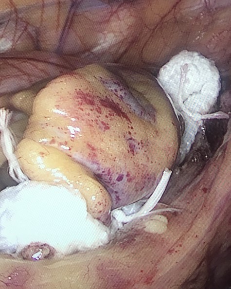
Atriclip™ in position at the base of the LAA.
Epicardial LAA clipping has been approved by the US-FDA. The AtriclipTM device (AtriclipTM Pro, AtriCure, Inc., Cincinnati, OH, USA) is one such device, and can be used to perform epicardial clipping of the LAA during both open and thoracoscopic surgery. Since its introduction in a prospective device trial in 2007 (NCT00567515), it has been shown to be safe, effective and durable in producing closure of the LAA [4]. No studies to our knowledge have examined the use of epicardial clipping as an adjunctive treatment in patients undergoing non-cardiac thoracic surgery with a diagnosis of AF. This case demonstrates the feasibility of such an approach as a concomitant procedure in a patient undergoing video-assisted thoracoscopic surgery (VATS) left upper lobectomy for a primary lung cancer.
CASE REPORT
A 70-year-old gentleman with a history of hypertension and type II Diabetes Mellitus presented with an incidental finding of a left upper lobe mass on chest radiograph. The lesion was fluorodeoxyglucose (FDG)-avid on PET-CT. While a CT-guided biopsy of the mass was non-diagnostic, a provisional diagnosis of a cT2aN0 non-small lung cancer was given. A left VATS upper lobectomy was planned. On attending the pre-operative assessment clinic, the patient was in AF with a pulse rate of 107, and a calculated CHA2DS2-VASc score of 2 (hypertension, diabetes mellitus). Apixaban was commenced, as well as bisoprolol for rate control. Pre-operative echocardiography was performed.
Concomitant atrial appendage clipping was performed at the time of the patient’s planned lobectomy. Access was via a standard anterior thoracoscopic approach using a three ports technique with the utility port anterior to latissimus dorsi in the fourth intercostal space, with camera and accessory ports in the sixth and seventh intercostal spaces. Thoracoscopic left upper lobectomy and systematic lymph node dissection was performed in a standard fashion. The authors do not consider there to be any advantage associated with epicardial clipping either pre- or post-lobectomy.
The AtriClipTM device (AtriCure, West Chester, OH, USA) is a self-closing rectangular device available in four sizes—35, 40, 45 and 50 mm. It can be placed thoracoscopically, through median sternotomy or thoracotomy. Intra-operative trans-oesophageal echocardiogram (TOE) was performed to out-rule thrombus within the LAA. The pericardium was opened by ~5 cm overlying the LAA, parallel and ~1 cm posterior to the phrenic nerve, using hook diathermy. The LAA neck interface with the true atrium was identified, along with the ligament of Marshall and the left circumflex coronary artery. The AtriClipTM device was placed through the inferior thoracoscopic port site incision, which had to be extended to ~2.5 cm to allow for device access. The 40-mm clip was guided over the LAA, and moved into position at the appendage base (Fig. 1). The clip was then closed and TOE used to confirm satisfactory occlusion. Once satisfied with the position, the clip was released from the guiding system and the guiding system withdrawn. The pericardial opening was not closed. A 24-Fr chest drain was placed prior to closure. Successful placement was defined on TOE as no residual flow to the LAA and a residual stump of <10 mm.
Post-operative histology confirmed a diagnosis of pT2aN0 adenocarcinoma. The patient remains in AF. At current follow-up, he has had no thromboembolic events, and is undergoing cardiology review to determine the risk-benefit of discontinuing anti-coagulation.
DISCUSSION
Epicardial clipping using the AtriClip™ device has consistently been shown to produce high rates of complete LAA closure, with success rates of >95% reported across a number of studies [5, 6, 7]. These studies also report exceptionally low rates of device-related complications, and low rates of post-operative bleeding and peri-operative stroke. Furthermore, the device can be placed at the time of surgery for other indications.
A number of studies have demonstrated the feasibility and safety of epicardial clipping by thoracoscopic approaches. Ellis et al.[6] reported a 93.85% success rate for AtriClip™ LAA closures performed thoracoscopically, with no post-operative thrombus or embolic strokes recorded over 183 patient-years. They highlighted the advantage of the short procedural time and the advantage that the AtriClipTM device has over devices such as the Watchman occlusion device in that placement does not mandate that the patient be on post-procedural anticoagulation [6]. Osmancik et al.[7], while acknowledging the additional challenge of thoracoscopic clip placement in comparison to open techniques, reported similar outcomes to studies in which the AtriclipTM was placed via median sternotomy. They recorded a successful occlusion rate of 97.5% across 40 patients, with once clip malplacement observed [7]. Similarly, Kurfirst et al.[5] published a case series of patients in whom the AtriClipTM was placed via both open and thoracoscopic methods. Fifty-six percent of 101 patients in this cohort underwent epicardial clipping by a thoracoscopic approach. Successful clip placement was achieved in 98% of patients with no clip migration, device leakage or post-operative appendage thrombus formation visualized on post-operative imaging (TOE and/or CT) [5]. In none of these studies were any incidences of ‘narrow-channel failure’—i.e. persistent flow to the LAA secondary to incomplete occlusion—recorded.
This case report highlights the feasibility of atrial appendage clipping in the setting of procedures being performed in the left hemithorax for indications other than AF—in this case, a primary lung malignancy. Further studies will no doubt examine the use of epicardial clipping in thoracic surgery to further establish the safety and efficacy of this emerging device.
Conflict of interest statement
None declared.
Sources of financial support
None.
