-
PDF
- Split View
-
Views
-
Cite
Cite
Andrew D Hart-Pinto, Rashid Sheikh, David Luff, Curious case of a calcified food bolus, Journal of Surgical Case Reports, Volume 2018, Issue 3, March 2018, rjy035, https://doi.org/10.1093/jscr/rjy035
Close - Share Icon Share
Abstract
Authors present an 80-year-old male attending with obstructive food bolus. Lateral soft tissue neck x-ray demonstrated a suspected calcified foreign body at the level of the larynx. Subsequent senior radiological input reported the findings as incidental calcification of carotid arteries. ENT surgeons should demonstrate increased awareness for potentially calcified soft tissues on interpreting such x-rays.
INTRODUCTION
Patients presenting to ENT services with impacted food bolus in itself is not an unusual phenomenon, with incidence approaching 13 in 100 000 [1]. Management of the condition involves directed history, endoscopic evaluation. Patients commonly complain of dysphagia, excessive salivation and sensation of foreign body in throat.
Radiographic soft tissue x-rays can provide advantageous information for the identification of radio-opaque foreign materials, e.g. fish or chicken bones. This can be invaluable amongst patients where primary observation and medical management has failed and surgical intervention becomes a requirement.
CASE REPORT
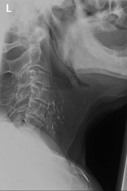
Authors present an 80-year-old male attending with symptoms of dysphagia, drooling and globus sensation. Symptoms commenced during the patients evening meal. The patient reported eating pork, with no recollection for presence of bones. The patient’s medical history was complicated by previous squamous cell carcinoma of the vocal cords requiring laryngectomy plus neck dissection 8 years previously. Despite multiple attempts to swallow the food bolus, supplemented by assistance from carbonated drinks, no improvement was observed. Doctors performed naso-endoscopic evaluation of the pharynx, and no visualization of the food bolus was noted. For completeness, a lateral soft tissue neck x-ray was performed. Radiographs demonstrated the presence of a calcified area within the larynx thought, initially by junior colleagues, to represent a foreign body (Fig. 1). The patient was placed upon regular hyoscine butylbromide injections, and observed until morning with a plan for rigid endoscopic evaluation plus removal of calcified piece of meat.

Lateral view soft tissue neck radiograph demonstrating calcified lesion at level of C4–C5. Presence of previous ligation clips noted from previous laryngectomy.
During morning ward round review; the senior consultant requested a repeat trial of oral liquids. Doctors were subsequently presented with a very pleased patient plus a self-evacuated food bolus. The food bolus demonstrated no evidence for the presence of bone, in correlation with the calcification observed on initial x-rays. A complete resolution of the patient’s symptoms was observed, being able to eat and drink without restriction. X-rays were reviewed by lead ENT and Radiology consultants. The ‘unusual foreign body’ is reported to be soft tissue calcification likely within the carotid artery.
DISCUSSION
Carotid artery arthrosclerosis has several risk factors including increasing age, smoking, dyslipidaemia and cardiovascular disease [2]. Amongst these patients, lateral soft tissue neck x-rays being used for identification of foreign bodies, increased awareness of calcification of surrounding soft tissues may mask the true presentation. Incidental calcification of carotid arteries have been previously reported as high as 5.06% in patients undergoing routine dental x-rays [3], therefore, appropriate translation is required on interpretation of lateral soft tissue neck x-rays.
Clinicians working up impacted food bolus patients should display vigilance in ensuring clinical history and endoscopic findings establish appropriate correlation with radiological findings.
LEARNING POINTS
Soft tissue x-rays are known to be helpful with identification of radio-opaque foreign bodies. They provide detailed information of the level at which the obstruction lies, permitting pre-operative planning in cases requiring surgical intervention.
Increased awareness amongst clinicians of potentially calcified soft tissues. In this case calcification present within the carotid artery was assumed to be the foreign body in question.
Importance of correlation of history and clinical findings with radiographic imaging. The most obvious answer is not always the correct one.
CONFLICT OF INTEREST STATEMENT
None declared.


