


Abstract
Superior patellar dislocation is a very rare pathology, which happens in middle age. We report a case of spontaneous superior patellar dislocation, which occurred at 30 years of age. Differential diagnosis is patellar tendon rupture, which could be reliably excluded by clinical examination and radiographs. Reduction manoeuvres should be tried with diluted intra-articular local anaesthetic for hydrodilatation to gain mechanical advantage. Splinting the knee in gentle flexion is recommended if general anaesthetic is employed. Recurrent dislocation or osteochondral fractures warrants surgical treatment.
Introduction
Because of its biomechanical properties, patellofemoral articulation is one of the most complex joints. Patella is suspended by tendons and stabilized by ligaments and is prone to dislocate if an imbalance arises [1]. Superior dislocation of patella is a rare condition with sixth decade being the peak age of presentation. In ~20 reported cases the youngest is a 43-year old [2]. We present a case of spontaneous superior dislocation of patella in a 30-year-old male with no obvious patellofemoral arthrosis.
Case Report
A 30-year-old gentleman woke up with a sudden onset of knee pain. He had never experienced any knee problems in past. The emergency department referred him as a suspected patellar tendon rupture. History, examination and radiographs revealed a characteristic superior patellar dislocation. No obvious generalized ligament laxity, genu recurvatum or patella alta was found. Hydrodilatation of his knee was performed using 10 ml of 0.5% Chirocaine diluted in 20 ml of 0.9% normal saline. Dislocation was reduced successfully with gentle digital manipulation. Full pain free range of motion was demonstrated after reduction. He was discharged home the same day with no recurrence at 6 months.
Discussion
Superior patellar dislocation is a rare pathology with patella alta, ligament laxity, neurological diseases and genu recurvatum being risk factors. Watson Jones reported first case of superior patellar dislocation in 1956 [3].
Common mechanism of injury is hyperextension of the knee associated with direct force to patella. Voluntary [4] and spontaneous [5, 6] superior patellar dislocations are also reported.
A thorough history and careful examination is essential for diagnosis. Due to its rarity, the emergency department usually refer this condition as patellar tendon rupture. It is easy to diagnose if clinician is aware and vigilant. Patient will not be able to do a straight leg raise in both conditions, but for different reasons. It is due to pain in superior patellar dislocation, while loss of extensor mechanism continuity is the cause in patellar tendon rupture. Most cases involved trivial or no trauma in superior patellar dislocation, with contraction of quadriceps in extended knee. Superior movement of the patella locks its lower articular margin on upper border of trochlea. The less frequent mechanism is direct impact on patella. In contrast, rupture of patellar tendon requires significant force and often is a consequence of eccentric contraction of quadriceps.
Knee cannot be flexed actively or passively in superior patellar dislocation, which is the most important differentiating point. Patients with patellar tendon rupture generally have less pain, knee can be passively flexed and often a gap is palpable in patellar tendon area. It should not be forgotten that a tense hematoma in tendon gap is possible.
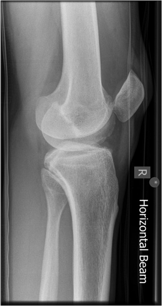
Lateral radiograph of knee showing dislocated patella.
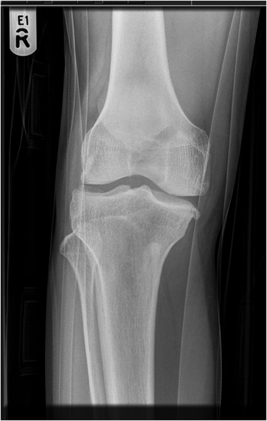
AP radiograph of knee.
Reduction can be attempted with intra-articular local anaesthetic, sedation, femoral nerve block and rarely general anaesthetic [7]. It is advised to immobilize knee in gentle flexion during recovery from general anaesthesia to avoid recurrence in perioperative period [4]. When awake, knee can be left free. Intra-articular local anaesthetic diluted in normal saline is recommended, giving analgesic as well as mechanical advantage, by pushing patella away from femur. This technique of getting a mechanical advantage has not been previously described in literature. If other analgesics fail, a general anaesthetic is used as a last resort and spontaneous reduction can happen [8]. In our experience, intra-articular diluted local anaesthetic provided very good pain relief. Spontaneous reduction of joint has also been reported after adequate analgesia [7].
The next important step is to hyperextend the knee as much as allowed comfortably by patient; this releases tension from patella and helps it unlock it from its position. Ipsilateral hip flexion with extended knee also helps to relax quadriceps further due to proximal attachment of rectus femoris. Two digits should be used under medial and lateral borders of patella to lift it away from surface of femur. Medial and lateral rocking movements of patella, while keeping it lifted, helps to relocate patella onto its track on trochlear groove. Once free, superior and inferior movement of the patella is felt and the knee can be fully flexed. Until the patella is unlocked, it is important not to crank the knee to flexion due to risk of iatrogenic injury to patellar tendon, inferior pole of patella or osteochondral fracture. Ability to freely flex knee actively and passively denotes relocation of patella. In all the reported cases, close reduction was successful except one [6]. Our alternate plan was to reduce the dislocation in general anaesthetic. If digital manipulation was unsuccessful, our plan was to use a pointed reduction clamp, after making small skin incisions to grasp patella, caudal to its equator, to manipulate. Effective lifting force can be applied through reduction clamp by eliminating slipping of skin and subcutaneous tissues over patella. The last option is to do arthroscopy. This has an advantage of shaving off the prominent osteophytes from inferior pole of patella and superior margin of femoral trochlea. No fractures of osteophytes have been reported in literature. The examiner should check for full range of movement, to detect the tendency to dislocate and feel for any crepitus. The presence of a crepitus may indicate osteochondral fracture.
Superior patellar dislocation is a rare problem. It can be treated by simple manoeuvres in accident and emergency avoiding unnecessary suffering and admission.
Acknowledgements
Authors gratefully acknowledge Hijab Fatima's (First Author's daughter) help in proofreading and improving presentation.
