


Abstract
A rare complication of diverticular disease is the formation of fistulas, most commonly either colo-vesical or colo-vaginal. We present the unusual case of a perforated sigmoid diverticulum forming a colo-gluteal fistula and presenting initially as a gluteal abscess in an otherwise asymptomatic patient. After drainage of the gluteal abscess, the patient re-presented with faecal loss from the abscess drainage site. Imaging revealed fistulous communication between the sigmoid and the left obturator internus muscle, tracking to the gluteus maximus with associated abscess and cutaneous communication to the site of previous drainage. The patient underwent an emergency Hartmann's procedure with lay open/abscess drainage of the gluteal cavity. Post-operatively the patient experienced continuing discharge from the gluteal fistula despite repeated drainage and debridement causing considerable morbidity, inconvenience and misery. Clinicians should maintain a high index of suspicion when presented with a gluteal abscess and should consider the possibility of an intra-abdominal source.
Introduction
Diverticular disease is a common condition with an increasing prevalence in western populations [1]. It affects patients mainly over the age of 40 and is associated with a number of risk factors including obesity, smoking and a low-fibre diet.
Clinical manifestations can range from mild/isolated symptoms to severe or recurrent disease. The commonest presentation is with acute left-sided abdominal pain; however, other presentations occur and are usually secondary to complications of the disease [2]. Such complications can include haemorrhage, stenosis, abscess formation, perforation, obstruction and or the formation of fistulae.
Fistulae disease are rare, occurring in ~12% of cases [3]. They usually form when a diverticular abscess/phlegmon ruptures into an adjacent structure and while theoretically any adjacent organ can be involved, the commonest fistulae are colo-vesical or colo-vaginal [4]. In contrast, colo-enteric, colo-uterine, colo-uretral and colo-cutaneous fistulas are rarer [5].
In this report, a case of fistulating sigmoid diverticulae resulting in a sigmoid-gluteal fistula and presenting initially as a peri-anal abscess in an otherwise asymptomatic patient is presented.
Case Report
A 67-year-old male was admitted to our acute unit with a 1-week history of a tender swelling over his left buttock/peri-anal margin. A course of oral antibiotics prescribed by his general practitioner had made no difference to his symptoms. He complained of severe pain in the left buttock associated with feverish symptoms. With hindsight, the pelvic/buttock pain seemed excessive compared with the outward appearance of the buttock swelling. He denied abdominal pain or alteration in bowel habit. His past medical history was significant for hypertension, atrial fibrillation, multiple previous myocardial infarctions and insertion of coronary stents. There was no past history of abdominal pathology.
Examination revealed a 2 cm area of tender erythema and induration over the left buttock/peri-anal region. There was also some bony tenderness noted on palpation of the left hip. Rectal examination was unremarkable.
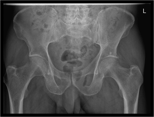
Pelvic X-ray showed a gas shadow over left obturator foramen.
The patient re-presented a week after discharge, complaining off persistent pain around the buttock, with feculent discharge and flatulence from the wound. Abscess cultures from the previous admission were reviewed and noted to have grown Escherichia coli and clostridium species.
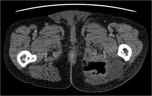
Enhanced CT showed an abscess in left gluteus maximus muscle.
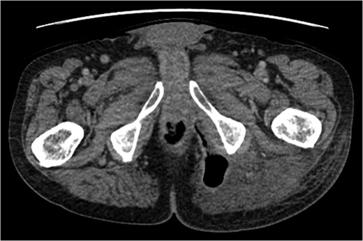
Fistula tracking to peri-anal margin.
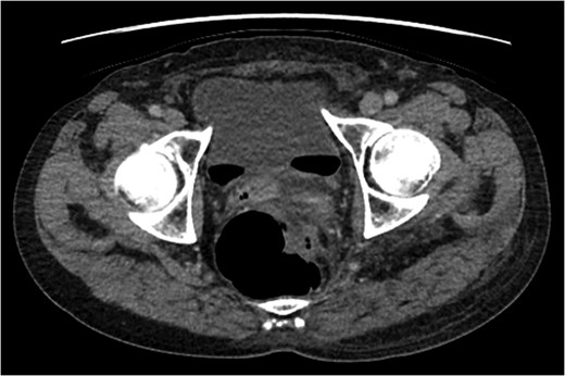
Evidence of colo-vesical fistula (free air in bladder).
An emergency Hartmann's procedure was performed under general anaesthetic through a lower midline incision as well as lay open of the perineal fistula. On opening the abdomen, a perforated diverticulum was identified extending into the left lateral pelvic wall. A standard Hartmann's procedure was performed, with sigmoid colectomy, division of rectum at recto-sigmoid junction and a tension-free stoma formed in left hypochondrium. The left buttock abscess cavity extended by a further 2–3 in. and the cavity then packed.
Following the operation, the patient had a short and uncomplicated critical care stay and was subsequently discharged. He has since continued to experience ongoing discharge from the fistula with follow-up imaging showing a reduction in the size of the abscess cavity but continuing patency of the fistula tract, despite repeated wound debridement and packing, drainage of the fistula and rectal washout.
Discussion
Fistula formation is a recognised, albeit infrequent, complication of diverticular disease. When this complication does arise, however, the fistulas are usually either colo-vesical or colo-vaginal in nature. In contrast, colo-cutaneous fistulas are much less common, accounting for only 1–4% of all diverticular fistulas [5].
The majority of colo-cutaneous fistulas reported in the literature are found communicating between the colon and the anterior abdominal wall and typically arise as a complication of drainage of abdominal abscess secondary to diverticular perforation, or spontaneous discharge of such abscess through skin [2, 4, 5].
To our knowledge, there are only a handful of reported cases where a diverticular perforation has formed a fistula to the gluteal region [6, 7]. The case that we describe is particularly rare given the absence of gastro-intestinal signs.
Peri-anal and buttock abscesses are common, patients usually have the abscesses drained by the junior member of the on call team, and commonly discharged without follow-up or reviewed in a telephone clinic. Patients who go on to develop signs of recurrent sepsis/fistulae-in-ano are then offered clinic appointments. It is not routine practice, therefore, for these common presenting conditions to be imaged, unless an index of suspicion is entertained.
The culture results in this case showing the presence of E. coli and clostridium species, along with flatulence and feculent discharge from the wound pointed towards a fistulous communication with the gastro-intestinal tract. These pointers were, however, realised after the patient had suffered considerable distress with symptoms and delay in diagnosis.
Despite considerable review of this case, the only pointers to the gravity of the presenting pathology was the degree of pain seeming to be excessive compared to the outward appearances of the abscess, and hip discomfort. It is not our routine practice to image all index acute presentations of peri-anal/buttock abscesses.
The published literature leaves us none the wiser and we recommend clinicians should be aware of this unusual presentation when presented with a gluteal abscess and should maintain a high index of suspicion and consider the possibility of an intra-abdominal source.
