-
PDF
- Split View
-
Views
-
Cite
Cite
Marco Yat Hang Yung, Jennifer Murray, Errington C. Thompson, Blunt aortic trauma in a patient with the Ehlers–Danlos syndrome type VI, Journal of Surgical Case Reports, Volume 2016, Issue 3, March 2016, rjw026, https://doi.org/10.1093/jscr/rjw026
Close - Share Icon Share
Abstract
A 24-year-old male with the Ehlers–Danlos syndrome (EDS) type VI (ocular scoliotic) who was kicked in the abdomen presented to the emergency room (ER) with abdominal pain. He was found to have a blunt traumatic aortic injury. The patient was treated nonoperatively. He was stable and discharged home on the eighth day. The patient returned to the ER several days later hypotensive and tachycardic. The patient was taken immediately to the operating room, but vascular repair was not possible. The patient expired. We discuss the challenges of taking care of a patient with EDS and offer suggestions that might improve future patient's outcome.
INTRODUCTION
The Ehlers–Danlos syndrome (EDS) is a connective tissue disorder characterized by hypermobility of the joints as well as hyperextensibility of the skin. EDS is not one disorder, but a group of disorders. Classic EDS (type I) is an autosomal dominant disorder with a defect in type V collagen. It is characterized by severe skin involvement. Hypermobility EDS (type III) is autosomal dominant and has a defect in tenascin X. These patients have classic severe joint hypermobility problems. Vascular EDS (also known as type IV) is an autosomal dominant disease, which is well known to the surgical community because of its propensity to cause spontaneous rupture of organs and arteries. In this variant, the defect is in collagen III. Ocular B scoliotic EDS (type VI) is known for severe curvature of the spine. Patients have a marfanoid-type body habitus. Because of the defect in procollagen B lysine 5 dioxygenase activity, minor trauma to the eye can cause globe rupture. These patients can also have rupture of the great vessels [1] (Table 1). To our knowledge, this is the first report of a traumatic dissection in a patient with EDS type VI. Because this patient was relatively well known in the medical community, Institutional Review Board approval was obtained for this case report.
| Type . | Typical features . | Inheritance . | Protein defect . |
|---|---|---|---|
| Classic (EDS I and II) | Skin hyperextensibility and fragility, joint hypermobility, tissue fragility manifested by widened hypertrophic scarring | Autosomal dominant | Collagen V |
| Hypermobile (EDS III) | Joint hypermobility, moderate skin involvement | Autosomal dominant | Tenascin X |
| Vascular (EDS IV) | Spontaneous rupture of internal organs including major arteries and intestines; skin is thin and translucent with extensive bruising; hypermobile minor joints | Autosomal dominant | Collagen III |
| X-linked EDS | Similar to classic type | X-linked recessive | Unknown |
| Ocular—scoliotic EDS VI | Muscular hypotonia, progressive kyphoscoliosis, marfanoid habitus, osteopenia, occasional rupture of eye globe and great vessels. Includes classic features of EDS | Autosomal recessive | Lysyl hydroxylase deficiency relative to prolyl hydroxylase activity |
CASE REPORT
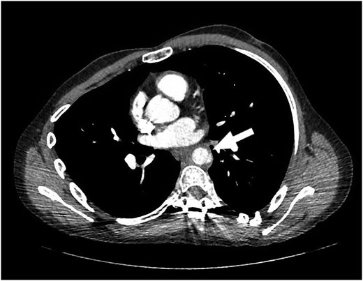
Our patient is a 24-year-old male with the EDS type VI who had undergone rod fixation of his spine for scoliosis, ocular surgery for a ruptured globe and ligation of his popliteal artery after an attempt was made to repair his aneurysm. The patient was being followed at Duke EDS Clinic where he underwent an extensive workup, and a definitive diagnosis was established. He presented to the emergency room (ER) after being kicked in the abdomen during a martial arts class. The patient felt an instant abdominal pain which resolved quickly. The patient went home after the incident and then developed nausea, vomiting and diaphoresis. He was hemodynamically normal in the ER. His groin pulses were slightly diminished compared with his radial pulses. The patient underwent a computed tomography (CT) scan of the chest, abdomen and pelvis with intravenous contrast. The patient was found to have traumatic aortic injury (Fig. 1). Cardiovascular surgery was consulted. The patient was admitted to the ICU for nonoperative therapy. He was started on Esmolol intravenously, and Diltiazem was added in order to keep his mean arterial pressure around 60. After 2 days in the ICU, the patient was transitioned to medications by mouth. He was allowed to ambulate on the fourth day and discharged on the eighth day after a long discussion with him and his family.

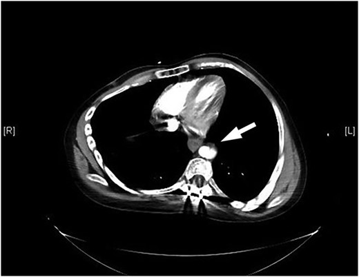
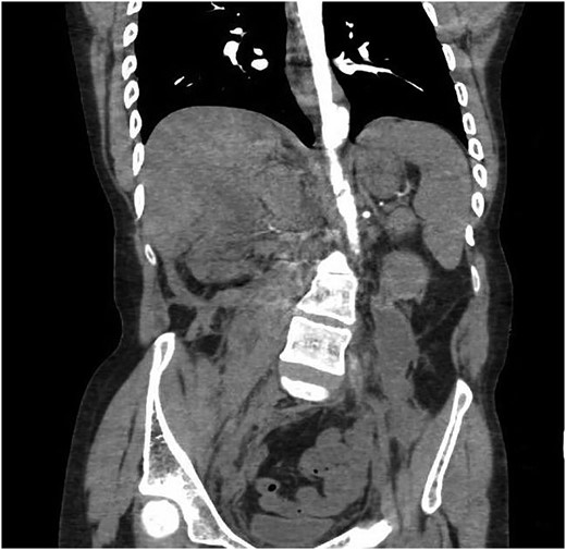
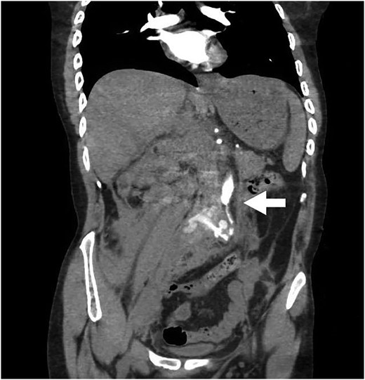
The patient returned to the hospital less than a week later in extremis. He had severe back pain and was diaphoretic. The patient was hypotensive and tachycardiac. He had no femoral pulses. Repeat CT scan revealed extension of the aortic injury in the abdominal aorta with no flow in the iliac (Figs 2–4). The patient was emergently taken to the operating room (OR) where the aorta, iliac and femoral vessels were unable to hold sutures. We were unable to repair this patient's aorta. Post-operatively, we spoke with the family, and the patient was allowed to expire.


Note contrast in the distal aorta but no contrast in the iliac arteries.
DISCUSSION
The EDS type VI has ocular, vascular and musculoskeletal manifestations. Complications of the disease continually plague patients throughout their lives. EDS type VI is caused by a defect in the lysyl hydroxylase enzyme [2].
Collagen metabolism is extremely complex and detailed [2]. Collagen is assembled from a single strand of procollagen into a triple helix. The N and C terminals are cleaved. Now, the collagen is grouped together in fibrils. Cross linking at the lysine and hydroxylysine is a key step in order for the fibrils to form. The fibrils then assemble into the strong collagen fibers [3]. The defect in the EDS type VI is reduced activity of the lysyl hydroxylase enzyme. This seems to lead to muscle hypotonia, joint laxity, kyphoscoliosis, poor wound healing, ocular fragility and arterial rupture [2].
The major problem in EDS is the patient's tissue-paper-like vessels. This makes it almost impossible for the vessels to hold suture. Some authors have suggested that pledgets should be used [4]. This can be helpful, but sometimes the tissue is too fragile for even pledgets. Because the vessels are so delicate, endovascular stents may be out of the question. All stents have some sort of anchor or hooks which hold the stents in place. These hooks would tear the brittle vessels in the EDS patient and therefore would not be useful. However, there have been a couple of recent case reports [5, 6] and one small retrospective series [7] suggesting that endovascular stents may have a role in selected patients with EDS.
In retrospect, it is hard not to look at this case and see that this patient had few viable options when he originally presented to the ER. Taking this patient to the OR was futile, as his vessels were unable to hold suture with or without pledgets. We suspect that this patient developed relative hypertension after discharge home. This caused more stress on his aorta through increased shearing forces. Because of the patient's inability to lay down normal collagen, he probably needed more time to heal this injury. Nonoperative therapy was the best option for the patient. Maybe keeping this patient in the hospital and making sure that his blood pressure and heart rate were under better control would have given this patient a better chance at survival.
CONFLICT OF INTEREST STATEMENT
None declared.


