


Abstract
Sebaceous naevus is a rare non-melanocytic congenital skin hamartoma. Even more rare is the transformation of these lesions into malignant skin cancers, most notably basal cell carcinomas (BCCs). We discuss a case in an adult with later malignant transformation into BCC reported by clinical pathologists. There is dispute about the accurate incidence of malignant transformation. More recently, research has shown that transformation into BCC is unlikely, in that the origins of these lesions arise in trichoblastoma. Evidence from this comes from the changes in mutation pathways that are distinctly separate. A consensus between dermatology, plastic surgery and clinical pathologists is beneficial to decide the best management for these individuals. The limiting factor is the agreement of diagnosis, compounded with conflicting large series multi-centre trials to determine the likelihood of transformation. We feel that each case should be managed individually taking into account patient and/or parent preference with the surgeon offering advice to help the patient guide management decisions. Patient/parent education is important and long-term follow-up by plastic surgeons or dermatologists is highly recommended.
INTRODUCTION
Sebaceous naevus (SN) is a non-melanocytic congenital skin hamartoma. Known as naevus sebaceum of Jadassohn, it was coined to describe a defective pilosebaceous unit. First described in 1895, it has a prevalence of 0.3% in neonates, with 95% of lesions located in the head and neck region [1]. Defined as hyperplasia of sebaceous glands, apocrine glands, hair follicles and epidermis, it presents as a pink raised verrucous plaque. There is a transition in tissue morphology from childhood to puberty and then again into adulthood. During infancy, they are flat lesions that develop into a raised wart-like lesion under the influence of hormones acting on the sebaceous glands. During puberty and adulthood, there have been documented cases of sebaceous naevi transforming into benign and malignant neoplasms [2]. Basal cell carcinoma (BCC) is a rare consequence of sebaceous naevi, and we discuss a case in an adult to highlight the need for continued vigilance.
CASE REPORT
A 60-year-old male was seen in the plastic surgery outpatient department with a right temporal lesion. The lesion had been present since birth but the patient never sought medical attention in the past since it remained asymptomatic. He decided to seek medical attention because the lesion had increased in size over a 5-year period, with intermittent symptoms of itchiness, bleeding and weeping. He was otherwise fit and healthy with no significant medical history other than well-controlled hypertension. He had no excessive prior exposure to ultraviolet radiation and was of Fitzpatrick skin type III.
Examination revealed a 40-by-30 mm fleshy verrucous lesion (Figure 1). There were no features suggestive of BCC (rolled edge, ulceration and telangiectasia). No other similar skin lesions elsewhere on his body were found on examination, nor any head and neck lymphadenopathy.
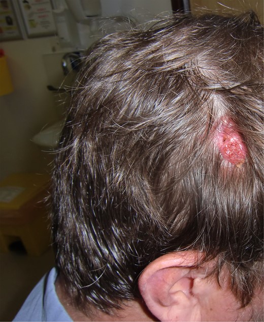
Sebaceous naevus right parietal. Typical waxy appearance within hair-bearing region.
An excision biopsy was performed under local anaesthetic with primary closure. Biopsy revealed a BCC, nodular type in an SN. Histology confirmed the lesion to be fully excised circumferentially at all margins with a minimum margin of 4 mm. There was invasion into the reticular dermis (5 mm) with no evidence of lymphovascular or perineural invasion. The specimen contained lymphoid aggregates, which were well circumscribed likely to represent pseudolymphoma (an inflammatory response with benign accumulation of inflammatory cells). The term pseudolymphoma is an umbrella term to describe an inflammatory infiltrate that resembles lymphoma but not diagnostic of the condition. It is most-often non-specific [3]. Interpretation of these cutaneous lymphoid aggregates has led to diagnostic confusion as they mimic lymphoid malignancy. However, the histological appearances require clinical history, examination and light microscopy findings to enable a firm diagnosis. However, there is no direct link that can be identified with SN or BCC. The patient remains well with no further evidence of recurrence at the excision site.
DISCUSSION
The lifetime risk for malignant change of SN has been suggested to vary from between 5 and 22% with BCC primarily the most likely malignancy [4]. There has been much discussion about the true incidence of BCC arising in sebaceous naevi. An 18-year review, suggested that the incidence was in the region of 0.8% (651 excised lesions) [5]. The authors believe serious consideration should be made to excise all sebaceous naevi. This was mainly due to the difficulties in identifying BCC in these lesions regardless of age group [6]. Others argue, however, that the cost in terms of morbidity and expense do not justify prophylactic excision. They suggest that patient education and monitoring of these lesions are sufficient, since BCC rarely metastasize. Concerning lesions should be referred to dermatology and plastic surgery colleagues for biopsy [5].
Furthermore, there has also been some debate for the optimal timing in excising these lesions. On-going debate continues in weighing up surgical excision against the potential for malignant transformation. A general consensus remains that lesions should be surgically excised if they exhibit suspicious features, as in our patient. A study evaluated 757 cases of SN in those aged under 16 and found no cases of BCC [7]. However, two cases of malignant transformation in sebaceous naevi have been described in children including squamous cell carcinoma, which has greater potential to metastasize, and also transformation into a microcystic adnexal carcinoma [8]. Therefore, it is also suggested that early excision before potential transformation during puberty should be considered [9].
Diagnostic confusion may be attributed to the wide range of true BCC in these lesions, which lie in the origins of trichoblastoma, a benign brown nodular lesion. This is grossly similar to BCC, and only differentiated on deeper evaluation histologically, with low mitotic rate, high in fibrocystic stroma with primitive follicular structures. Therefore, it has been contended that BCC does not arise from an SN, but merely these tumours are trichoblastomas. This has been supported in genetic studies. RAS mutations are found whereby malignant transformation occurs within an SN. When a BCC arises in SN, this typically shows Hedgehog pathway dysregulation, not RAS mutations [10].
While there is no definitive management plan for patients with sebaceous naevi, we know these lesions have malignant potential. There is a divided opinion on the incidence and lifetime risk of malignant transformation. However, this appears to be low lifetime risk. We feel therefore that each case should be managed individually taking into account patient and/or parent preference with the surgeon offering advice to help the patient guide management decisions. Patient/parent education is important and long-term follow-up by plastic surgeons or dermatologists is highly recommended.
CONFLICT OF INTEREST STATEMENT
None declared.
