


Abstract
We present a case of a 61-year-old male presenting with post-prandial epigastric pain and marked weight loss. Investigation revealed calcific atherosclerosis of the abdominal aorta, coeliac axis, superior mesenteric (SMA) and renal arteries. He had undergone radiotherapy for testicular teratoma 34 years previously. Percutaneous mesenteric revascularization by primary stenting of the SMA proved successful. Radiotherapy for intra-abdominal malignancy has the potential to induce both acute and chronic enteritis and an accelerated atherosclerotic process in the arteries within the field of beam.
INTRODUCTION
Radiotherapy for the treatment of malignancy is a common practice and the continuous optimization of this treatment raises the problem of long-term issues of patients treated and cured by ionizing radiation [1]. Accelerated atherosclerosis secondary to radiation arteritis has been previously described in the literature in up to 40% of patients [1–3].
The site of treatment determines the arteries at risk of damage in the irradiated field; for example, coronary artery disease and arterial occlusive disease after radiotherapy to the chest following treatment for lymphoma or breast cancer [2] and carotid artery disease following head and neck radiotherapy [1]. A variety of complications have been described in the literature in relation to abdominal and pelvic irradiation including enteritis, mesenteric ischaemia and renal artery stenosis [4].
Although testicular cancer is a common malignancy, with the mainstay of treatment being orchidectomy, lymph node dissection and adjunctive radiotherapy, descriptions of vascular complications following treatment for these germ cell tumours are limited in the literature [3, 5].
CASE REPORT
A 61-year-old male was referred to the Department of Vascular Surgery from the gastroenterology service with a 2-year history of post-prandial abdominal pain, particularly when eating larger meals which evolved to a disinclination to eat and total weight loss of three stones.
His previous medical history included a right orchidectomy followed by a 4-week course of abdominal radiotherapy for testicular teratoma in 1978, irritable bowel syndrome, sleep apnoea, hypothyroidism, hypertension and sinus node dysfunction. He was not diabetic and had never smoked. Physical examination revealed signs of weight loss but no other stigmata of disease.
Gastroscopy revealed a benign duodenal stricture between D1 and D2, which subsequently resolved on treatment. An abdominal CT scan identified a lesion in the right lobe of the liver deemed benign on further CT assessment with triple-phase contrast. However, the latter also revealed heavy calcification throughout the abdominal aorta with severe stenosis at the origins of the coeliac axis, bilateral renal, superior mesenteric and inferior mesenteric arteries (Fig. 1).
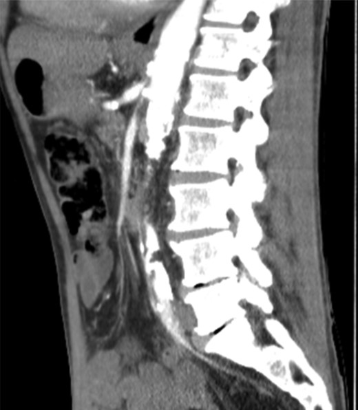
CT angiography—heavy calcification through abdominal aorta and severe stenosis of coeliac axis, renal arteries, SMA and IMA.
Tortuous vessels collateralizing with the intrahepatic arteries had developed to supply the liver via the diaphragmatic branches to compensate for impaired blood flow in the common hepatic artery. A vasculitis screen was negative.
Following discussion at the vascular multi-disciplinary team meeting successful mesenteric revascularization was performed. The ostium of SMA was accessed using a SIM-1 catheter; angiography confirmed a tight stenosis of the SMA, which was transversed using a Terumo wire and 6-French destination sheath after angioplasty with a 3 × 20 mm balloon. A 6 × 18 mm balloon with expandable stent completely restored the calibre of the vessel with good antegrade flow and resolution of symptoms.
DISCUSSION
Radiotherapy for intra-abdominal malignancy has the potential to induce both acute and chronic enteritis and an accelerated atherosclerotic process in the arteries within the field of beam [4, 6]. Radiation-induced arterial disease differs from naturally occurring atherosclerosis and is thought to be due to a process of subendothelial connective tissue proliferation, disruption of elastic lamina, fibrosis of adventitia, obliteration of the vasa vasorum and reduced bioavailability of nitric oxide in irradiated tissue [6].
The lesions are localized to the field of radiation and manifestation of multi-organ atherosclerosis is often lacking [1, 2, 5, 6]. The chronic effects of irradiation damage to arteries do not present acutely but can develop insidiously over a longer term; hence, the association with abdominal or vascular symptoms may be delayed in many cases [1, 3, 4]. This is supported by cases of radiation damage to arteries at other sites including enteritis following treatment for Wilms tumour [6], stroke after head and neck radiotherapy [2], ischaemic heart disease and subclavian stenosis following radiotherapy for lymphoma and breast cancer [1, 5] and intermittent claudication following external iliac stenosis after radiotherapy for cervical and testicular cancer [5].
Our patient did not have classical risk factors for atherosclerosis, which was only localized to the field of the external beam radiotherapy. Endovascular management with angioplasty and stent was the treatment modality of choice which has given a good short-term result. The literature has described the comparative results of open versus endovascular mesenteric revascularization [7–10]. The benefits of an endovascular procedure include a quicker procedure with lower perioperative mortality rates and a shorter hospital stay. Long-term patency is improved in those undergoing open repair but surgery within a previously irradiated field can be difficult with distorted anatomy, increased risk of bleeding and longer healing times. Despite this, long-term survival rates appear comparable in the two groups [7, 8].
Recent epidemiological studies have shown that radiation-induced accelerated atherosclerosis is underestimated with no real prospective studies available [1]. The management of these patients with or without other vascular risk factors remains to be determined as the exact pathophysiological mechanism of arterial damage is uncertain. Prolonged vascular follow-up for this at-risk group of patients seems pertinent together with the use of preventive strategies including antiplatelet agents and statins. As survival rates and treatment of cancer improves it may be that this delayed consequence of radiotherapy becomes an increasing clinical problem.
