Journal Article

A painless swelling of the abdominal wall

Journal of Surgical Case Reports, Volume 2014, Issue 3, March 2014, rju011, https://doi.org/10.1093/jscr/rju011
Published:
05 March 2014
Article history
Received:
06 December 2013
Accepted:
29 January 2014
Published:
05 March 2014

Abstract

Case report. A man with a painless swelling of the abdominal wall. A 77-year-old male presented with a progressively increasing painless swelling of the abdominal wall due to nodular fasciitis.

INTRODUCTION

A general physician refers a 77-year-old male with no previous illnesses to the department of surgery. At the time of presentation a subcostal swelling of the abdominal wall has been present for over 5 months. This painless swelling has progressively grown in size over the past few months.

CASE REPORT

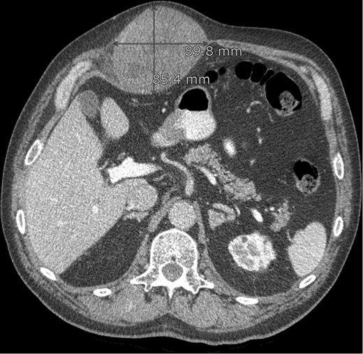
On palpation the lesion is solid elastic to the touch. An ultrasound investigation with histological biopsy was made. Histological examination showed fibroma myxoid tissue with the suspicion on nodular fasciitis. Additional CAT-scan images (Fig. 1) revealed a smooth, oval and the peritoneum-bordered tumour located medially and spreading into the rectus abdominis muscle.
CT abdomen: lesion in the abdominal wall, possibly originating from the rectus abdominis muscle.
Figure 1:

CT abdomen: lesion in the abdominal wall, possibly originating from the rectus abdominis muscle.

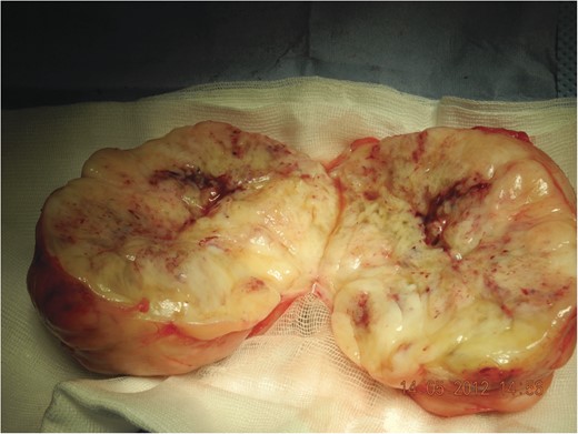
During surgery a large yellow lobular partially fibrotic mass (Fig. 2) was excised located entirely above the muscle fascia. Histological examination showed nodular fasciitis without signs of malignancy.
Tumour ex vivo, note the lobular structure with fibrous septa (incised).
Figure 2:

Tumour ex vivo, note the lobular structure with fibrous septa (incised).

DISCUSSION

Nodular fasciitis, also known as pseudosarcomatous fibromatosis, is predominately seen in young adults (20–40 years). Mostly affecting the upper extremities or trunk it can mimic a malignant sarcoma due to its rapid growth.

This is an Open Access article distributed under the terms of the Creative Commons Attribution Non-Commercial License (http://creativecommons.org/licenses/by-nc/3.0/), which permits non-commercial re-use, distribution, and reproduction in any medium, provided the original work is properly cited. For commercial re-use, please contact journals.permissions@oup.com

Supplementary data

Close
This Feature Is Available To Subscribers Only

Sign In or Create an Account

Close

This PDF is available to Subscribers Only

View Article Abstract & Purchase Options

For full access to this pdf, sign in to an existing account, or purchase an annual subscription.

Close

Gift article access

As a benefit of your subscription, you can share temporary access to restricted articles.

Each link will stop working after 30 days or 10 uses. You may create up to 10 links in a 30 day period.

Please sign in to your personal account to gift article access.

Gift article access

Please create a link below to share with others. This will provide temporary access to this restricted article.

The link will stop working after 30 days or 10 uses. You may create up to 10 links in a 30 day period.

Gift articles remaining: --

Gift article access

The link will stop working after 30 days or 10 uses. You may create up to 10 links in a 30 day period.

Gift articles remaining: --

Gift article access

As a benefit of your subscription, you can share temporary access to restricted articles.

Each link will stop working after 30 days or 10 uses.

You have reached the limit of 10 links within a 30 day period