


Abstract
We report a case of recurrent severe upper gastrointestinal bleeding where the bleeding source was difficult to find during recurrent hospitalizations. Eventually videocapsule endoscopy was the modality that finally diagnosed an ulcerated lipoma within an area of intussuscepted jejunum. Segmental resection of small bowel was performed and no further bleeding episodes have occurred. Our case illustrates the value of capsule endoscopy and the rare potential of lipomas to cause serious gastrointestinal bleeding.
INTRODUCTION
Jejunal lipomas are rare benign submucosal tumors and often incidental findings. Their clinical behavior is usually silent, but occasionally acute gastrointestinal bleeding or intestinal obstruction may occur [1].
We present a patient with recurrent upper gastrointestinal bleeding who underwent repeated endoscopies followed by computed tomography and videocapsule endoscopy (VCE). This case demonstrates the value of the VCE in obscure GI bleeding.
CASE REPORT
A 53-year-old male presented to the emergency room with melena and severe anemia. During the past 6 months, the patient had four episodes of melena. Repeated gastroscopy and colonoscopy revealed no significant pathology. Past medical history included ischemic heart disease, congestive heart failure, insulin-dependent diabetes mellitus, chronic renal failure, peripheral vascular disease and COPD.
The laboratory tests showed a hemoglobin level of 6.5 mg/dl, urea 130 mg/dl and creatinine of 3.5 mg/dl. The patient was treated conservatively. An isotopic study with Tc-99 did not reveal any bleeding source. Computed tomography was initially interpreted as normal. A VCE study was performed that identified a submucosal bulge with a central ulceration in the proximal third of the small bowel (Fig. 1). More distally in the small intestine melena could be seen by the capsule (Fig. 2). Revision of the CT scan confirmed the VCE finding and showed a lesion of fat density in jejunum.
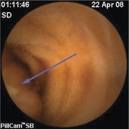
Videocapsule endoscopy image of a submucosal bulge with a superficial ulceration (arrow) in the proximal small intestine.
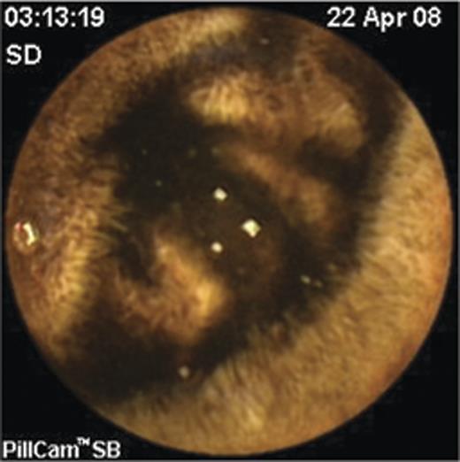
Videocapsule endoscopy image of melena within a more distal small bowel loop.
The patient was referred to surgery and exploratory laparatomy diagnosed intussusception of jejunum containing an intraluminal submucosal lesion of 5 cm with a superficial ulceration (Fig. 3). Segmental resection of the jejunum was performed. Histology confirmed the submucosal lesion to be a lipoma. The postoperative course was uneventful and the patient was discharged on the 6th postoperative day.
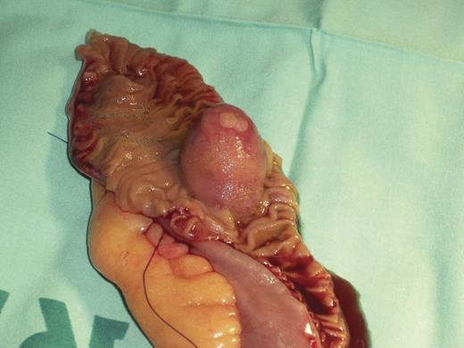
Surgical specimen of a short segment of resected jejunum showing the lipoma with a small superficial ulceration.
DISCUSSION
Lipomas of the gastrointestinal tract are rare especially in the jejunum. The most frequent site is the large intestine where they occur in 65–75% of cases followed by the stomach in 20–25% [2]. There is a small female preponderance and most cases appear in the 6th–7th decades of life. In the small bowel, lipomas are solitary in most cases. When large they rarely present with upper GI bleeding, intussusception and occlusion [1–7]. The GI bleeding caused by a large lipoma may be chronic as in our case. The bleeding is caused by ulceration of the mucosa due to mass enlargement coupled by normal peristalsis or direct pressure of the lipoma. Recently, two similar cases of long-standing occult GI bleeding caused by a small bowel lipoma were reported [6]. Both patients were diagnosed by the VCE after negative small bowel follow through studies. In both cases, a superficial ulceration was seen on the surface of the lipoma. Before these new technologies were available, diagnosis was often made only at laparatomy or intraoperative enteroscopy [8].
Overt upper GI bleeding rarely complicates large small bowel lipomas with a reported incidence of about 1% [8]. Diagnosing the bleeding source by angiography or radionuclide scan can be difficult due to the intermittent or low rate of bleeding. The VCE is capable, in most patients, of screening the entire small intestine and localizing the bleeding source to the proximal, middle or distal third of the small bowel based on the transit time of the capsule.
In our patient, the bleeding source was correctly diagnosed by the VCE. The unique VCE image (Fig. 1) of the jejunal lipoma with a superficial ulceration correlated surprisingly well with the finding that was eventually seen within the resected surgical specimen (Fig. 3). This case also supports the use of VCE as the initial modality for investigation of obscure gastrointestinal bleeding even in the hospital setup after ambulatory investigation failed due to problems of compliance.
