


Abstract
A pelvic digit is a rare congenital anomaly and often an incidental radiographic finding. Here we present the first reported case of a 57-year-old Caucasian female with a pelvic digit and a chief complaint of dyspareunia. Radiographic studies and magnetic resonance imaging confirmed a stable bony excrescence from the inferior left ischium impinging on the pelvic floor. The patient underwent surgical removal of the pelvic digit without complications. Accurate diagnosis of pelvic lesions is necessary to guide treatment.
INTRODUCTION
Pelvic digits are rare congenital anomalies that develop in soft tissue.(1-4) There are less than twenty reported cases in the literature. The majority of reported pelvic digits are asymptomatic and found incidentally during radiography.(2) To date, there has never been a report describing a patient with a pelvic digit who presents with a chief complaint of dyspareunia. To the best of our knowledge, this is the third reported case of a symptomatic pelvic digit and emphasizes the importance of considering lesions of the bony pelvis in the differential diagnosis of dyspareunia.
CASE REPORT
A 57-year-old Caucasian female presented with a chief complaint of more than one year of dyspareunia. She also experienced pain with ambulation and now regularly used a walker. The pain was localized to the lateral aspect of her perineum. She did not have any history of pelvic trauma or radiation. Her past medical history includes chronic bilateral knee, hip and ankle pain and had a right total hip arthroplasty that required multiple revisions.
Physical examination revealed a prominence in the perineal area that was tender to palpation. She had no pain with hip range of motion and her motor and sensory examinations were intact.
Review of her radiographs demonstrated a well-positioned right total hip arthroplasty without any evidence of loosening or wear. Her left hip was unremarkable. However, there was an osseous growth projecting from the inferior aspect of left ischium toward the perineal region. Additional imaging with computed tomography (CT) and non contrast magnetic resonance (MR) were used to further define the lesion.
Representative images, including: a plain radiograph (Figure 1), 3D reconstructed computed tomography (Figue 2) and T1 coronal magnetic resonance imaging (Figure 3) indicated a bony excrescence from the inferior aspect of the left ischium measuring 1.7 x 1.1 cm. MR without gadolinium did not show any surrounding inflammatory changes. No aggressive features were seen on the studies. Review of a previous CT scan from 2 years prior to the most recent study, demonstrated a stable lesion.
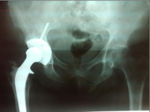
Plain X-ray: AP pelvis demonstrating pelvic digit arising from the left ischium
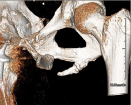
3D Reconstructed Computed Tomography demonstrating the pelvic digit
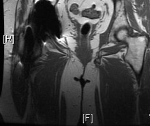
Magnetic Resonance Imaging demonstrating a pelvic digit
The above studies were suggestive of a pelvic digit. The patient’s chief complaint was painful intercourse and she elected to proceed with a resection. A pelvic digit was further confirmed after resection (Figure 4). Pathology found no evidence of malignancy and the patient was discharged without pain and able to ambulate without support of a walker. The removed pelvic digit had two bony segments that formed a pseudoarticulartion or joint, visible in images A and B. Follow up via a telephone communication indicated that her dyspareunia had resolved.
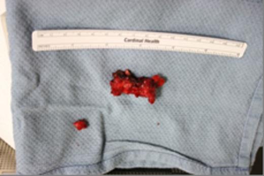
Surgical specimen of the removed pelvic digit
DISCUSSION
Pelvic digits remain an anomaly. Literature reports have suggested various origins of the pelvic digit, including congenital formation, myositis ossificans, or traumatic avulsion.(1-3,5,6) In this case, the defining features were that it was well corticated with a pseudoarticulation between bony segments, resembling a digit. This is most consistent with a congenital etiology. Typically this would occur prior to the sixth week of fetal life in the mesynchymal stage.(6,8)
In most reported cases the pelvic digit is an incidental finding on radiography. There are only two other cases of symptomatic pelvic digits reported in the literature. Pandey et al. and Maegele report the only other cases of symptomatic pelvic digits in a 35 and a 40 year old man, respectively.(6,9) These patients both experienced chronic hip pain, tenderness to palpation of the affected region and limited range of motion.(6,9) Surgical excision was curative for chronic pain and increasing range of motion in both instances.(6,9)
In summary, pelvic digits are typically incidental findings on imaging studies. However, in order to facilitate the diagnosis and treatment of pelvic conditions, physicians should be aware that this anomaly can be a source of pain and disability.
