


Abstract
Pilonidal sinus is a common condition that typically affects the sacrococcygeal region of young men, whereas involvement of other sites, including the external genitalia, is exceedingly rare. We report the case of a 22-year-old man with a penile pilonidal sinus who had failed to respond to multiple courses of antibiotic therapy. Physical examination revealed a firm white papule with a central pinpoint opening on the dorsal balanopreputial fold. The lesion was surgically excised, and histopathological examination confirmed the diagnosis of pilonidal sinus. The postoperative course was uneventful, with complete healing and no complications. Although rare, penile pilonidal sinus should be considered in the differential diagnosis of suspicious penile lesions, alongside both benign and malignant conditions.
Introduction
Pilonidal sinus is a chronic inflammatory condition that most commonly affects the natal cleft of young men, while involvement of other anatomical sites is rare [1]. Unusual locations include the umbilicus, suprapubic region, external genitalia, axilla, sternum, breast, intermammary area, and interdigital clefts [2]. We report the case of a 22-year-old man with a penile pilonidal sinus successfully treated by radical circumcision, achieving an excellent functional outcome. Diagnosis is usually clinical, and advanced imaging is rarely required. The differential diagnosis includes balanoposthitis, hidradenitis suppurativa, epidermoid cysts, follicular occlusion disorders, and penile carcinoma. Careful clinical assessment and histopathological examination are essential for accurate diagnosis.
Case presentation
A 22-year-old male was referred urgently to our Urology Unit for evaluation of a long-standing suspicious penile lesion that had been progressively increasing in size and was associated with localized inflammation. He had previously undergone several courses of antibiotic therapy, with no significant improvement. The patient had no relevant past medical history.
On clinical examination, a white, indurated papule with a central pinpoint orifice was identified on the dorsal aspect of the balanopreputial fold (Fig. 1). No palpable inguinal lymphadenopathy was noted. During surgery, the lesion was found to be densely adherent to the surrounding tissues, including the tunica albuginea and the penile neurovascular bundle. A radical sleeve circumcision with penile reconstruction was performed, and the excised foreskin was sent for histopathological analysis. Histopathological examination revealed a terminal hair embedded within the epidermis, accompanied by multinucleated giant cells in the underlying dermis (Fig. 2a). Free hair shafts were seen traversing the inflammatory focus (Fig. 2b), surrounded by a dense lymphocytic infiltrate.
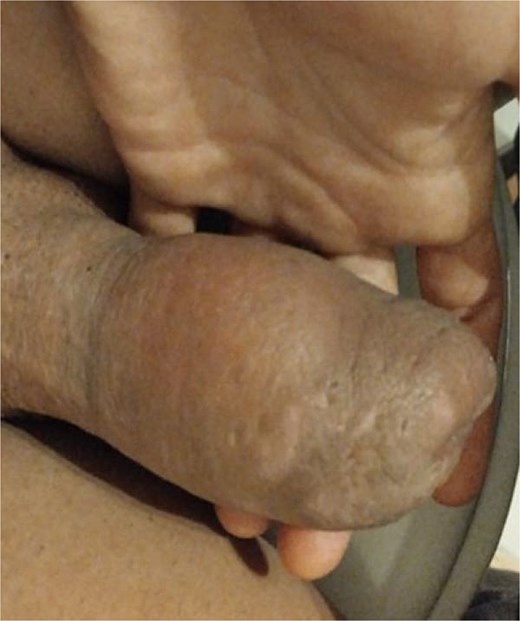
Picture of penile pilonidal sinus: swelling at the distal penile shaft.
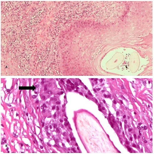
Histopathological examination of the specimen (Hematoxylin and Eosin stain). (a) Pilonidal sinus with areas of chronic inflammation and degenerative hair shaft (40×). (b) At high magnification hair shafts are seen in the center of this pilonidal sinus, surrounded by inflammation with foreign body giant cells (arrow) (400×).
The patient had an uneventful postoperative recovery, and there was no recurrence of symptoms during follow-up.
Discussion
Pilonidal sinus most commonly affects the sacrococcygeal region; however, rare cases have been reported in other locations, including the umbilicus, suprapubic area, external genitalia, axilla, sternum, breast, intermammary area, and interdigital clefts [2–4].
The etiology of pilonidal sinus disease remains controversial. The most widely accepted theory considers it an acquired condition in which hairs penetrate the skin due to local mechanical forces and friction, leading to follicular inflammation, infection, and abscess formation [5, 6].
Alternatively, some authors have proposed that the hairs found in pilonidal sinuses may not originate from the surrounding skin but are instead shed hairs from other areas that become trapped within a developing subcutaneous cavity [7, 8]. Patey and Scarff [7] demonstrated that loose hairs can penetrate the skin through minor defects, while Doll et al. [8] highlighted the role of environmental factors in hair accumulation.
Additional risk factors include a sedentary lifestyle, elevated body mass index, and hirsutism. Histologically, the disease is characterized by a sinus tract extending into the subcutaneous tissue. Follicular occlusion by keratin leads to inflammation, rupture, and formation of a pilonidal microabscess.
A pilonidal sinus of the penis (PSD) is a rare clinical entity, with only 29 cases reported in the literature to date [9–11]. A summary of these cases is provided in Table 1. The reported patient ages range from 19 to 61 years. In these individual case reports, no consistent occupational or comorbid risk factors were identified. However, some authors have speculated on a possible association between PSD and self-neglect in patients with psychiatric illness, although the evidence remains inconclusive [11].
Summary of documented cases in the medical literature.
| References | Age | Symptoms | Site |
|---|---|---|---|
| Yates-Bell (1968) | 29 | S A | Dorsal |
| Bervar et al. (1968) | N/A | N/A | N/A |
| Eckhart (1969) | N/A | N/A | N/A |
| Ritchie (1975) | 59 | D P | Dorsal |
| Fisher et al. (1976) | 43 | P A | Dorsal |
| Goudarzi and McColl (1976) | 40 | S P | Dorsal |
| Griffin et al. (1990) | 29 | U | Corona of penis |
| Rashid et al. (1992) | 21 | Dy | Corona of penis |
| Khan and Scott (1992) | 46 | A S | Dorsal |
| Burgess et al. (1992) | 24 | A U | Dorsal |
| Lingam et al. (1996) | 30 | P A | Ventral |
| Saharay et al. (1997) | 53 | S | Ventral |
| Val-Bernal et al. (1999) | 25 | P A | Dorsal |
| Val-Bernal et al. (1999) | 28 | S | Dorsal |
| O’Kane et al. (2004) | 61 | U | Ventral |
| Chikkamuniyappa et al. (2004) | 42 | S P | Beneath foreskin |
| Chikkamuniyappa et al. (2004) | 22 | D-P | Dorsal |
| Kalsi et al. (2004) | 56 | A | Dorsolateral base of glans |
| Rao et al. (2006) | 21 | S | Coronal sulcus |
| Al Chalabi et al. (2008) | 24 | U | Dorsal mid shaft |
| Sion-Vardy et al. (2009) | 41 | N/A | Midshaft |
| Goulao et al. (2009) | 30 | S | Dorsal glans |
| Al-Qassim et al. (2013) | 19 | D S | Dorsal distal shaft |
| Cormio et al. (2013) | 26 | U | Dorsal gland |
| Navarrete et al. (2017) | 20 | Dy | Dorsal balanopreputial fold |
| Ercil et al. (2018) | 20 | D | Dorsal midshaft |
| Shanmugathas N et al. (2020) | 28 | U | Coronal sulcus |
| Keurlings PAJ (2022) | 43 | N/A | N/A |
| Sawaqed F et al. (2025) | 30 | S | Mid-shaft |
S, swelling; A, abscesses; D, discharge; P, phimosis; U, ulceration; Dy, dyspareunia; N/A, not applicable.
Clinical presentation varies and may include abscess formation, purulent discharge, dyspareunia, penile swelling, phimosis, and rarely erectile dysfunction [9]. Lesions are most commonly located on the dorsal aspect of the uncircumcised penis near the coronal sulcus, which may act as a site for accumulation of hairs and debris. Notably, twelve published cases did not report the presence or absence of inguinal lymphadenopathy, despite its relevance in distinguishing PSD from malignant lesions [1, 12].
Most cases were managed surgically through excision of the sinus tract, often combined with circumcision. Histopathological examination, performed in nearly all reported cases, remains the diagnostic gold standard [9]. Although PSD is usually diagnosed clinically, untreated lesions may progress to chronic sinus formation containing retained hairs. Histology typically demonstrates chronic inflammation with foreign-body giant cells and embedded hair shafts, distinguishing PSD from true cystic lesions [11].
PSD is typically diagnosed clinically, with features resembling abscesses in other anatomical regions. If untreated, a chronic pilonidal sinus may develop, often containing retained hairs within the cavity or sinus tract. Depending on the severity of the condition, various surgical treatment options are available, including incision and drainage of the abscess and excision of the sinus tract. In most cases, the wound is allowed to heal by secondary intention [13].
Although often mischaracterized as cysts, pilonidal sinuses lack a true epithelial lining. Histopathological analysis usually reveals chronic inflammation with foreign body giant cells and embedded hair shafts, distinguishing PSD from other cystic lesions [14].
Complications include secondary bacterial or fungal infection and, rarely, malignant transformation into squamous cell carcinoma [15]. Definitive treatment consists of complete excision followed by primary closure or healing by secondary intention. In the present case, excisional biopsy with primary closure achieved satisfactory healing without functional impairment. Given its ability to mimic both benign and malignant penile lesions, PSD should be considered in the differential diagnosis of penile masses to facilitate timely management and reduce morbidity.
Conflicts of interest
The authors declare no conflicts of interest.
Funding
This work received no external funding.
Informed consent
Informed consent was taken from the patient for the publication of this case report.


