


 https://orcid.org/0009-0000-4082-4838

Abstract
Hepatic mesenchymal hamartoma (HMH) is a rare benign paediatric liver tumour. We present an 8-month-old female infant referred with a 2-month history of progressive abdominal distension and poor oral intake. Contrast-enhanced computed tomography identified a massive heterogeneous right hepatic mass measuring 10.7 × 14 × 15.4 cm, extending to the pelvis, crossing the midline, and abutting major vascular structures. Ultrasound-guided Tru-cut biopsy confirmed HMH. Following multidisciplinary team (MDT) discussion, open right hepatectomy was performed achieving complete tumour excision with clear surgical margins of 7 mm. Histopathology confirmed a benign biphasic mesenchymal–biliary proliferation without atypia or malignant transformation. The patient had an uneventful postoperative recovery with preserved liver function. Radiological follow-up at 4 months demonstrated a clear operative bed with no residual or recurrent disease. This case highlights the diagnostic and surgical challenges of giant HMH and the critical role of an MDT approach in achieving curative resection.
Introduction
Hepatic mesenchymal hamartoma (HMH) is an uncommon benign developmental lesion of the liver, representing the second most frequent benign hepatic tumour of childhood. It accounts for ~8% of all paediatric liver tumours and is characterized by an anomalous proliferation of primitive hepatic mesenchyme, giving rise to a biphasic composition of loosely arranged myxoid stroma interspersed with bile duct structures and islands of entrapped hepatocytes [1, 2]. The majority of cases manifest within the first 2 years of life, with a slight male predominance, although presentation at any paediatric age has been documented [3].
Clinically, HMH typically presents with progressive abdominal distension caused by a rapidly enlarging hepatic mass. In large or giant lesions, mechanical compression of adjacent structures may produce secondary symptoms including respiratory distress, compromised oral intake, and urological effects. Biochemical markers such as alpha-fetoprotein (AFP) may be mildly elevated, further complicating differentiation from malignant entities such as hepatoblastoma and undifferentiated embryonal sarcoma (UES) [4, 5].
Imaging, while highly informative, does not always yield a definitive diagnosis owing to the heterogeneous radiological spectrum of HMH, ranging from predominantly cystic to predominantly solid lesions. Contrast-enhanced computed tomography (CECT) and magnetic resonance imaging (MRI) are essential for characterizing the lesion, defining its relationship to vascular structures, and planning surgical resection [6]. Pre-operative biopsy may be required in diagnostically challenging cases.
Complete surgical resection remains the cornerstone of management, eliminating the risk of rare but documented malignant transformation into UES. For large or anatomically complex lesions, formal hepatectomy under multidisciplinary team (MDT) guidance is frequently necessary [7, 8]. We herein report the case of an 8-month-old Iraqi female infant presenting with a giant HMH measuring over 15 cm, which extended to the pelvis and abutted major vascular structures, successfully managed by open right hepatectomy following MDT discussion.
Case report
An 8-month-old female infant was referred to our tertiary paediatric surgical unit in Iraq with a 2-month history of progressive abdominal distension and poor oral intake. Caregivers reported gradual abdominal enlargement without associated fever, vomiting, jaundice, or pain. There was no significant birth history, dysmorphic features, or family history of hepatic disease.
On general examination, the infant was alert and conscious, in no apparent distress, with no pallor or jaundice. Abdominal examination revealed marked distension with a massive, ill-defined, non-tender mass occupying the majority of the abdominal cavity.
Abdominal ultrasound demonstrated hepatomegaly extending to the right iliac fossa, with a large heterogeneous, predominantly isoechoic mass measuring ~8.1 × 6.9 cm within the right hepatic lobe. The lesion exhibited internal cystic changes and exerted significant mass effect on adjacent vascular structures, with mild dilatation of the intrahepatic biliary tree.
CECT of the chest, abdomen, and pelvis revealed a right-sided abdominal mass measuring 10.7 × 14 × 15.4 cm, most likely arising from the liver, demonstrating heterogeneous enhancement with internal cystic degeneration. Superiorly, it abutted the main portal vein. Inferiorly, it extended into the pelvis to the level of S3, compressing the bladder dome. Medially, it crossed the midline, displacing the pancreas and bowel loops to the left. Posteriorly, it compressed the right kidney and abutted the aorta, inferior vena cava, and iliac vessels.
MRI of the abdomen, performed with coronal and sagittal T2-HASTE and post-contrast T1 sequences, confirmed a large exophytic heterogeneous mass with multilocular internal cystic components arising from the inferior aspect of the right hepatic lobe. The solid component demonstrated homogeneous moderate contrast enhancement, while the cystic components showed no enhancement. Right hepatic vessels were seen crossing into the lesion (Fig. 1).
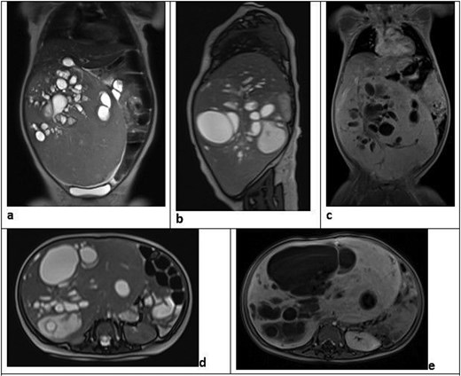
Pre-operative MRI of the abdomen. Coronal T2-HASTE (a), sagittal T2-HASTE (b), coronal post-contrast T1 (c), axial T2-HASTE (d), and axial post-contrast T1 (e) sequences demonstrate a large heterogeneous partially cystic, partially solid exophytic mass arising from the inferior aspect of the right hepatic lobe with multilocular internal cystic components. The cystic component shows no contrast enhancement, while the solid component demonstrates homogeneous moderate enhancement. Right hepatic vessels are seen crossing into the lesion.
Given diagnostic uncertainty, a percutaneous ultrasound-guided Tru-cut core needle biopsy was performed. Histopathological examination revealed features consistent with HMH: lobulated islands of expanded mesenchymal stroma containing loose connective tissue and epithelial bile ducts in varying proportions, with a loose myxoid background populated by bland spindle-shaped fibroblasts and dilated vascular channels. The biliary structures were tortuous and branched, resembling a ductal plate malformation pattern (Fig. 2, panels 1 and 2). Islands of entrapped hepatocytes with retained cell plate architecture were identified (Fig. 2, panel 3), alongside mesenchymal areas of bland spindle cells in loose stroma (Fig. 2, panel 4) and focal cystic degeneration with haemosiderin deposition (Fig. 2, panel 5).
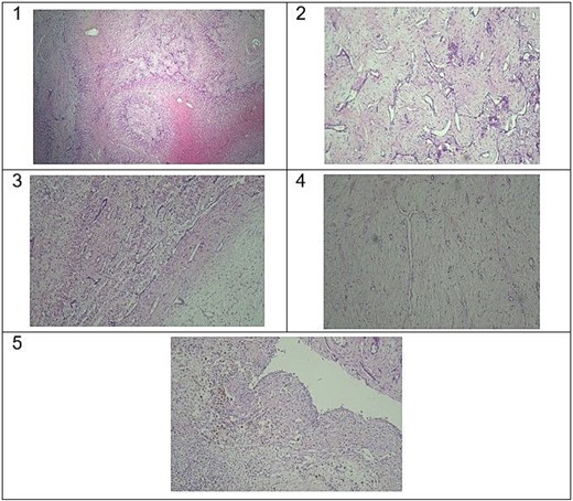
Histopathological features of hepatic mesenchymal hamartoma. Panel 1: loss of normal hepatic lobular architecture and absence of portal tracts [haematoxylin and eosin (H&E), ×4]. Panel 2: bland spindle cell myxoid stroma with collagen, containing branching and dilated bile ducts without cytological atypia (H&E, ×10). Panel 3: entrapped hepatocytes with retention of normal cell plate architecture (H&E, ×10). Panel 4: mesenchymal areas of bland spindle cells in loose stroma (H&E, ×10). Panel 5: areas of cystic degeneration with haemosiderin deposition (H&E, ×10).
Immunohistochemistry demonstrated diffuse moderate positivity for actin and desmin within the mesenchymal cells. Hepatic parenchymal cells showed a normal pericentral pattern of glypican-3 expression and membranous beta-catenin staining. CK19 highlighted the bile duct structures, while SALL-4 was negative, effectively excluding a germ cell or embryonal component.
The case was reviewed at a formal MDT meeting including paediatric surgeons, radiologists, histopathologists, and paediatric oncologists. Given the confirmed diagnosis of HMH, the massive size of the lesion, its extensive local mass effect, and the risk of malignant transformation, the consensus was to proceed with surgical resection.
The patient underwent open right hepatectomy via a bilateral subcostal (rooftop) incision under general anaesthesia. Intraoperative findings confirmed a large right hepatic tumour consistent with pre-operative imaging. The right hepatic artery and right portal vein were ligated and divided; right hepatic vein division and parenchymal transection were completed with careful haemostatic technique, achieving complete excision.
Postoperative histopathological examination of the resected specimen confirmed complete excision with clear surgical margins of ~7 mm, free of tumour. Microscopically, the lesion demonstrated a benign biphasic proliferation of loose myxoid mesenchymal stroma without cytological atypia, containing irregularly distributed bile ducts, cystically dilated spaces lined by flattened to cuboidal epithelium, and islands of entrapped hepatocytes. No cytological atypia, necrosis, or increased mitotic activity was observed.
The patient tolerated the procedure well. Vital signs remained stable; liver function tests and coagulation profiles were within normal limits throughout hospitalization. Pain was effectively managed, oral intake resumed gradually, and wound healing was satisfactory. The patient was discharged in good clinical condition.
Radiological follow-up on 29 March 2026 with axial T2 HASTE-FS and post-contrast T1 VIBE-DIXON MRI sequences demonstrated a clear operative bed with no residual enhancing lesions or postoperative collections, and no evidence of residual or recurrent disease (Fig. 3). The patient is currently under regular outpatient follow-up and remains well.
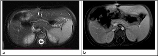
Post-operative MRI follow-up (29 March 2026). Axial T2 HASTE-FS (a) and post-contrast T1 VIBE-DIXON (b) sequences demonstrating a clear operative bed with no residual enhancing lesions or postoperative collections, and no evidence of residual or recurrent hepatic disease.
Discussion
HMH represents a significant diagnostic and surgical challenge, particularly when presenting as a giant mass in infancy. The present case is notable for several reasons: the massive tumour size exceeding 15 cm in a child aged only 8 months, the extensive anatomical involvement including pelvic extension and proximity to major vascular structures, and the successful achievement of complete resection with clear margins following a structured MDT approach.
The pathogenesis of HMH remains incompletely understood. Current evidence supports a developmental anomaly of primitive hepatic mesenchyme, possibly related to androgenetic-biparental mosaicism and chromosomal aberrations at the 19q13.4 locus, leading to activation of the chromosome 19 microRNA cluster (C19MC) [9]. This molecular background is shared with undifferentiated embryonal sarcoma of the liver, supporting the well-documented potential for malignant transformation and further justifying the rationale for complete surgical excision [10].
HMH most commonly presents in children under 2 years of age with progressive abdominal distension, as exemplified by this case. The absence of constitutional symptoms and negative AFP may assist in distinguishing HMH from hepatoblastoma, although AFP elevation has been reported in a minority of HMH cases and should not serve as a definitive discriminator [5, 11]. The differential diagnosis in this case included hepatoblastoma, infantile haemangioendothelioma, hydatid cyst, lymphangioma, and cystic teratoma, each of which was systematically excluded [6].
MRI demonstrated characteristic features: a large exophytic heterogeneous mass with multilocular cystic components arising from the right hepatic lobe, with moderate homogeneous enhancement of the solid component and absence of enhancement in cystic areas. This mixed cystic-stromal imaging pattern is well recognized in HMH, and the extraordinary extent—pelvic extension to S3, midline crossing, and portal vein abutment—underscored the critical importance of high-quality cross-sectional imaging in pre-operative planning [3, 6].
In diagnostically uncertain cases, pre-operative core needle biopsy provides indispensable histological characterization. Biopsy findings in this case demonstrated the hallmark biphasic morphology of HMH: loose myxoid mesenchymal stroma with bland spindle cells, tortuous bile ducts in a ductal plate malformation pattern, and islands of entrapped hepatocytes [1, 2]. The immunohistochemical profile—mesenchymal reactivity for actin and desmin, pericentral glypican-3 expression, membranous beta-catenin, CK19-positive bile ducts, and SALL-4 negativity—effectively excluded hepatoblastoma and embryonal tumours, providing diagnostic confidence prior to definitive surgery [2, 12].
Complete surgical resection is the definitive treatment for HMH, eliminating recurrence risk and the potential for malignant transformation [7, 8]. Open right hepatectomy was chosen over non-anatomical resection given the tumour’s size, segmental distribution, and intimate relationship with right-sided hepatic inflow and outflow structures. Clear surgical margins of 7 mm were achieved—a critical outcome, as incomplete resection has been associated with local recurrence [7, 13]. The uneventful postoperative course with preserved liver function and early enteral feeding is consistent with contemporary series reporting low morbidity following formal hepatic resection for HMH in experienced centres [8, 14].
Radiological follow-up at four months confirmed a disease-free operative bed, though long-term surveillance is recommended given the documented potential for late recurrence or malignant evolution [10, 15]. This case, together with our previously published experience [8], reinforces key lessons: the value of pre-operative biopsy in ambiguous presentations; the indispensable role of MRI in operative planning; the necessity of formal MDT discussion; and the curative potential of open hepatectomy even in anatomically challenging infant presentations. For centres in resource-limited settings, open hepatectomy remains a highly effective approach where laparoscopic facilities may be unavailable.
Acknowledgements
The authors thank the nursing, anaesthetic, and theatre teams at Warith International Cancer Institute for their dedicated contributions to the care of this patient.
Conflicts of interest
None declared.
Funding
No funding was received for this work.
Consent
Written informed consent was obtained from the patient’s legal guardian for the anonymized publication of clinical details, imaging, and histopathological images in this article. Our institution does not require separate ethical approval for the reporting of individual case reports.


