


 https://orcid.org/0009-0005-5350-2834

Abstract
Gastric volvulus with perforation is a rare and potentially catastrophic complication of hiatal hernia. Gastric volvulus complicates ~4% of hiatal hernia cases. We present a case of a 71-year-old male patient who presented with severe progressive generalized abdominal pain associated with absolute constipation, nausea, and vomiting ongoing for 8 days before admission. An abdominal computed tomography scan demonstrated a large hiatal hernia with organoaxial gastric volvulus and pneumoperitoneum around the stomach, denoting gastric volvulus with perforation. During laparotomy, the abdomen was found to be filled with turbid fluid, and the stomach herniated into the thoracic cavity, forming a gastric volvulus with signs of inflammation and a pyogenic membrane. There was a small peripyloric perforation ~5 mm. Repair of the hiatal hernia and gastric perforation was done. In this case report, we present the diagnostic approach and surgical options of a complicated hiatal hernia with gastric volvulus and perforation.
Introduction
A hiatal hernia is defined as the herniation of the gastric cardia and intra-abdominal content into the thorax due to the laxity of the esophageal hiatus in the diaphragm. Hiatal hernia is classified into four types. Type I, the sliding hiatal hernia, is the most commonly presented type, in which the gastroesophageal junction (GEJ) herniates into the thorax. Type II, the paraesophageal hernia, in which a portion of the stomach herniates into the thorax while the GEJ stays in its native site. Type III, Both the esophageal hiatus and a portion of the stomach herniate into the thorax. Type IV, where any portion of the stomach with additional abdominal viscera herniates into the thorax [1]. Hiatal hernias are mostly asymptomatic, especially in elderly patients, with an endoscopic detection rate of 0.8%–2.9% [2]. Nevertheless, symptoms vary according to the type of hiatal hernia; Type I presents with gastroesophageal reflux disease due to the loss of resting tone of the lower esophageal sphincter. While the other types of hiatal hernias can present with epigastric pain, nausea, vomiting, postprandial fullness, and dyspnea. Additionally, hiatal hernia symptoms can vary according to the associated complications, including gastritis, gastric ulceration, gastric volvulus, obstruction, incarceration, strangulation, necrosis, and perforation [3, 4].
Case report
A 71-year-old male patient with no significant medical or surgical history. Presented to the emergency department complaining of severe progressive generalized abdominal pain associated with absolute constipation, nausea, and vomiting, which had been ongoing for 8 days before admission. There were no previous episodes of similar abdominal pain. Upon admission, the patient appeared ill, and vital signs were as follows: blood pressure 146/110 mmHg, heart rate 96 bpm, oxygen saturation 93%, and temperature 37°C. On abdominal examination, abdominal distension was noted with generalized abdominal tenderness and localized guarding mainly at the epigastric area. Chest examination revealed equal bilateral air entry. Laboratory tests revealed leukocytosis with a white blood cell count of 31.22 × 10⁹/L, hypokalemia with a potassium level of 3.34 mEq/L, and hypomagnesemia with a magnesium level of 0.7 mEq/L. Venous blood gas showed mixed metabolic and respiratory acidosis with pH: 7.21, PaCO₂: 47 mmHg, and HCO₃−: 16 mmol/L.
An abdominal computed tomography (CT) scan demonstrated a large hiatal hernia within the thoracic cavity, with organoaxial gastric volvulus. Pneumoperitoneum was noted around the stomach, denoting gastric perforation, with extensive mesenteric fat stranding and free fluid seen at the gastrocolic ligament. Additionally, there was an abnormal configuration of the large bowel with cecum positioning in the right subphrenic space, denoting malrotation (Fig. 1).
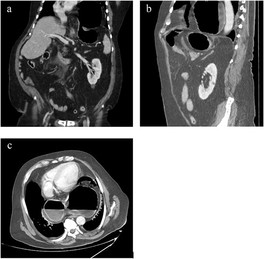
CT finding. (a) CT scan of abdomen, coronal view. (b) CT scan of abdomen, sagittal view. (c) CT scan of abdomen, axial view. Showing a large hiatal hernia with organoaxial gastric volvulus. Pneumoperitoneum was noted around the stomach, denoting gastric perforation, with extensive mesenteric fat stranding and free fluid at the gastrocolic ligament.
The patient was shifted from the emergency department to the operating room immediately and underwent laparotomy. During which, the abdomen was found to be filled with turbid fluid, and the stomach was herniated into the thoracic cavity, forming a gastric volvulus with signs of inflammation and a pyogenic membrane. There was a small peripyloric gastric perforation ~5 mm. The herniated stomach was reduced in the abdomen with sharp and blunt dissection, with elongation of the esophagus. Repair of the hiatal hernia with a figure-of-8 suture and repair of the peripyloric perforation with mattress suture were done using polydioxanone suture material. A nasogastric tube was inserted and placed in the duodenum. Additionally, copious washing with normal saline and drain insertion were done. Finally, closure of the abdominal wall with two loop sutures and closure of the skin with a skin stapler were done.
Seven days postoperatively, the patient developed wound dehiscence and was re-operated on, where a fascial defect was noted in the epigastric area, ~5 cm, and was closed with tension-free closure. The patient gradually improved and was discharged home after 27 days postoperatively.
Discussion
Gastric volvulus complicates ~4% of hiatal hernia cases [5]. It is defined as the rotation of the stomach or part of it by >180° around one of its axes [6].
Gastric volvulus is classified into four types according to the axis of rotation. Type 1, organoaxial volvulus, is the most commonly presented type, accounting for ~60% of cases, and includes the rotation of the stomach around the pylorus-cardia axis. Type 2, mesenteroaxial volvulus, includes the rotation of the stomach around an axis that bisects the greater and lesser curvatures. Type 3, which accounts for 2% of cases, is a combination of the first two types. Type 4 accounts for 10% of cases, which is an unclassified type [7].
The diagnostic approach includes comprehensive blood tests, cardiac enzymes, blood gases with lactate, and electrocardiogram to rule out other differential diagnoses of epigastric pain apart from the rare entity of complicated hiatal hernia. Chest X-rays can provide initial insights, including hiatal hernia, pneumoperitoneum, and pleural effusion, typically left-sided. An abdominal CT scan provides the definitive diagnosis and clarifies the associated complications [8].
Urgent surgical intervention for hiatal hernia is considered rare and required in only 1.16% of the cases, and it is associated with a high risk of mortality with a rate of 5%–17% [2]. Surgical decision-making and determining the appropriate surgical technique depend on several factors, including the patient’s condition, comorbidities, and risk of complications. Along with the visibility, perforation site, and intrathoracic invasion, which can be difficult to assess based on the CT scan images [2].
Thoracotomy exhibits more complications, including esophageal shortening and anastomotic dehiscence, although it provides better visibility and permits mediastinal lavage when indicated. In contrast, laparotomy is associated with limited visibility but fewer complications and facilitates applying omental patches while repairing the perforation [9, 10].
In a stable patient with a less complicated hiatal hernia, minimally invasive laparoscopic surgery can be considered. It provides better visibility around the esophageal hiatus but can be challenging in complicated cases that are associated with adhesions, inflammation, and hematomas [2].
In conclusion, this case highlights the diagnostic and management approach of a complicated hiatal hernia with gastric volvulus and perforation. Emphasizing the importance of early detection and surgical intervention to avoid the devastating complications and the associated surgical difficulties.
Conflicts of interest
None of the authors have any conflicts of interest to disclose concerning this article.
Funding
This research did not receive any specific grant from funding agencies in the public, commercial, or not-for-profit sectors.


