


 https://orcid.org/0000-0002-2947-1346

Abstract
Pyogenic granuloma (PG) is a benign vascular lesion that commonly occurs in the oral cavity during pregnancy, whereas cutaneous involvement of the hand is rare. I report a 33-year-old gravida 3 para 2 who presented at 39 weeks of gestation with a rapidly enlarging palmar mass measuring ~3 cm. The lesion became friable and interfered with hand function. Magnetic resonance imaging demonstrated a well-defined exophytic lesion confined to the superficial tissues without deep involvement. Surgical excision was performed four weeks postpartum under local anesthesia with sedation. Histopathological examination confirmed lobular capillary hemangioma with focal ulceration, granulation tissue, and reactive epithelial changes; immunohistochemistry for HHV-8 was negative. The postoperative course was uneventful, and no recurrence was observed at eight-week follow-up.
Introduction
Pyogenic granuloma (PG), also known as lobular capillary hemangioma, is a benign acquired vascular lesion characterized by rapid growth, friability, and a tendency to bleed [1]. It is frequently associated with local irritation or trauma and is influenced by hormonal changes, particularly during pregnancy, where it is commonly referred to as granuloma gravidarum [1, 2]. Oral lesions represent the most frequent presentation and have been reported in ~1.7%–8.5% of pregnancies [2, 3].
Cutaneous PG is less common and may occur at extraoral sites including the trunk and extremities [1]. Involvement of the hand is rare but clinically important because of its impact on daily function and the increased risk of bleeding. Unlike oral lesions, which may regress after delivery, cutaneous lesions are less likely to resolve spontaneously and often require surgical excision when symptomatic [4].
In atypical locations, PG may mimic malignant or aggressive vascular lesions, making histopathological examination essential for definitive diagnosis [5].
This report describes a rare case of palmar PG arising late in pregnancy and highlights diagnostic considerations and postpartum surgical excision as a safe and effective management strategy.
Case report
A 33-year-old gravida 3 para 2 woman with no known chronic illnesses or drug allergies presented with a progressively enlarging lesion on the palm of her left hand. The lesion initially appeared as a small papule approximately six months prior to presentation and gradually increased in size during pregnancy. By 39 weeks and 2 days of gestation, it measured ~3 × 3 cm and was associated with occasional discharge. The lesion became increasingly friable in late pregnancy, with bleeding on minimal trauma, resulting in functional limitation of the hand.
On examination at term, there was a red-brown, ulcerated, exophytic nodule located between the second and third metacarpals. There was no surrounding erythema, no signs of infection, and no regional lymphadenopathy. Hand perfusion, sensation, and range of motion were preserved.
The patient delivered a healthy infant by spontaneous vaginal delivery on 7 February 2025 without complications. Estimated blood loss was 200 mL, and a first-degree perineal tear was repaired. The postpartum period was uneventful.
Magnetic resonance imaging (MRI) performed on 13 February 2025 demonstrated a well-defined exophytic soft tissue lesion arising from the dermal and subdermal layers, measuring ~2 × 2 × 1.4 cm. The lesion showed a central vascular stalk with high signal intensity on T2-weighted images and low signal intensity on T1-weighted images. Mild surrounding subcutaneous edema and minimal tenosynovitis of the second flexor tendon were noted. The lesion was clearly separate from the underlying flexor tendons.
Surgical excision was performed four weeks postpartum under local anesthesia with sedation. The lesion was well circumscribed and attached by a narrow stalk, which was ligated and excised in the subcutaneous plane. Hemostasis was achieved without difficulty, and the specimen was sent for histopathological analysis.
Histopathological examination confirmed lobular capillary hemangioma (PG), demonstrating lobulated capillary proliferation with focal ulceration, granulation tissue, and reactive epithelial changes. Immunohistochemistry for HHV-8 was negative.
The postoperative course was uneventful. At eight-week follow-up, the surgical site was well healed, hand function was fully preserved, and there was no evidence of recurrence (Fig. 1).
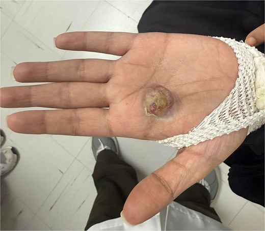
Palmar pyogenic granuloma (lobular capillary hemangioma) presenting as an ulcerated exophytic nodule between the second and third metacarpals.
Discussion
This case describes an uncommon presentation of PG involving the palm during late pregnancy. Cutaneous involvement of PG during pregnancy is less common than oral presentation, and lesions of the hand are rarely reported [3, 4]. When present on the palm, these lesions may significantly impair hand function and are prone to recurrent bleeding due to constant mechanical stress.
The pathogenesis of PG is multifactorial, with hormonal changes during pregnancy promoting angiogenesis and vascular proliferation, while local irritation or repeated trauma may further stimulate lesion development [1, 2]. In contrast to oral lesions, which may regress postpartum, cutaneous lesions are less likely to resolve and more often require surgical intervention when associated with symptoms or functional limitation [4].
In atypical anatomical locations, PG may clinically resemble malignant lesions, and histopathological confirmation is required to establish the diagnosis [5]. Imaging is not routinely necessary for typical lesions but may be useful in selected cases to assess lesion extent and exclude deeper involvement, particularly when clinical features are atypical [6].
In this patient, the lesion demonstrated progressive enlargement and increased friability during late pregnancy, leading to functional impairment. MRI confirmed that the lesion was confined to superficial tissues and was not involving deeper structures, which facilitated safe surgical planning.
Several features of this case distinguish it from previously reported pregnancy-associated hand lesions. The lesion attained an unusually large size of ~3 cm by term, with documented functional impairment severe enough that the patient was unable to use the hand normally. Furthermore, preoperative imaging raised hemangiosarcoma and other mesenchymal malignancies as genuine differential diagnoses, underscoring that clinical and radiological features alone may be insufficient to exclude malignancy in atypical palmar lesions during pregnancy. Notably, adequate surgical planning was achieved with non-contrast MRI, which may be preferable when contrast administration is a concern in the postpartum period.
The timing of intervention in pregnancy-associated PG should be individualized. In this case, excision was deferred until the postpartum period, allowing for improved hemostasis and safer operative conditions. Complete excision, including the lesion base, is important to minimize the risk of recurrence. At short-term follow-up, there was no evidence of recurrence, and full hand function was preserved.
Author contributions
The author was responsible for patient management, data collection, literature review, manuscript preparation, and final approval of the report.
Conflicts of interest
The author declares no competing interests.
Funding
No funding was received for the preparation of this manuscript.
Ethical approval
Ethical approval was not required for this case report in accordance with institutional guidelines.
Patient consent
Written informed consent was obtained from the patient for the publication of this case report and any accompanying images.


